 Movie
Movie Controller

[English] 日本語
 Yorodumi
- PDB-1byr: CRYSTAL STRUCTURE OF A PHOSPHOLIPASE D FAMILY MEMBER, NUC FROM SA... -
+ Open data

- Basic information
| Entry | Database: PDB / ID: 1byr | ||||||
|---|---|---|---|---|---|---|---|
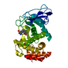
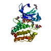
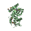
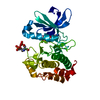
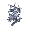
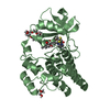
| Title | CRYSTAL STRUCTURE OF A PHOSPHOLIPASE D FAMILY MEMBER, NUC FROM SALMONELLA TYPHIMURIUM | ||||||
 Components Components | PROTEIN (ENDONUCLEASE) | ||||||
 Keywords Keywords | ENDONUCLEASE / PHOSPHODIESTERASE | ||||||
| Function / homology |  Function and homology information Function and homology informationphosphorus metabolic process / RNA endonuclease activity producing 5'-phosphomonoesters, hydrolytic mechanism / phospholipase D / D-type glycerophospholipase activity / lipid catabolic process Similarity search - Function | ||||||
| Biological species |  Salmonella typhimurium (bacteria) Salmonella typhimurium (bacteria) | ||||||
| Method |  X-RAY DIFFRACTION / MAD / Resolution: 2 Å X-RAY DIFFRACTION / MAD / Resolution: 2 Å | ||||||
 Authors Authors | Stuckey, J.A. / Dixon, J.E. | ||||||
 Citation Citation | Journal: Nat.Struct.Biol. / Year: 1999 Title: Crystal structure of a phospholipase D family member. Authors: Stuckey, J.A. / Dixon, J.E. | ||||||
| History |
|
- Structure visualization
| Structure viewer | Molecule: MolmilJmol/JSmol |
|---|
- Downloads & links
-Download
| PDBx/mmCIF format | 1byr.cif.gz | 45.9 KB | Display | PDBx/mmCIF format |
|---|---|---|---|---|
| PDB format | pdb1byr.ent.gz | 32.4 KB | Display | PDB format |
| PDBx/mmJSON format | 1byr.json.gz | Tree view | PDBx/mmJSON format | |
| Others |  Other downloads Other downloads |
-Validation report
| Arichive directory | https://data.pdbj.org/pub/pdb/validation_reports/by/1byrftp://data.pdbj.org/pub/pdb/validation_reports/by/1byr | HTTPS FTP |
|---|
-Related structure data


-Links
 PDBj
PDBj- Assembly
| Deposited unit | 
| ||||||||
|---|---|---|---|---|---|---|---|---|---|
| 1 | 
| ||||||||
| Unit cell |
|
-Components
| #1: Protein | Mass: 17175.416 Da / Num. of mol.: 1 Source method: isolated from a genetically manipulated source Source: (gene. exp.) Salmonella typhimurium (bacteria) / Cellular location: PERIPLASM / Gene: NUC / Plasmid: PT7-7 / Species (production host): Escherichia coli / Cellular location (production host): PERIPLASM / Gene (production host): NUC / Production host: |
|---|---|
| #2: Water | ChemComp-HOH /  Mass: 18.015 Da / Num. of mol.: 188 / Source method: isolated from a natural source / Formula: H2O Mass: 18.015 Da / Num. of mol.: 188 / Source method: isolated from a natural source / Formula: H2O |
-Experimental details
-Experiment
| Experiment | Method: X-RAY DIFFRACTION / Number of used crystals: 1 |
|---|
- Sample preparation
| Crystal | Density Matthews: 2.86 Å3/Da / Density % sol: 55 % | ||||||||||||||||||||
|---|---|---|---|---|---|---|---|---|---|---|---|---|---|---|---|---|---|---|---|---|---|
| Crystal grow | pH: 7.5 Details: 10 MG/ML PROTEIN + 2M NH4SO4, 100MM TRIS-HCL,PH 7.5 | ||||||||||||||||||||
| Crystal | *PLUS | ||||||||||||||||||||
| Crystal grow | *PLUS Method: vapor diffusion, hanging drop / Details: Zhao, Y., (1997) Protein Sci., 6, 2655. | ||||||||||||||||||||
| Components of the solutions | *PLUS
|
-Data collection
| Diffraction | Mean temperature: 123 K |
|---|---|
| Diffraction source | Source: ROTATING ANODE / Type: RIGAKU RU200 / Wavelength: 1.5418 |
| Detector | Type: ADSC |
| Radiation | Monochromator: NI FILTER / Protocol: SINGLE WAVELENGTH / Monochromatic (M) / Laue (L): M / Scattering type: x-ray |
| Radiation wavelength | Wavelength: 1.5418 Å / Relative weight: 1 |
| Reflection | Resolution: 2→16.4 Å / Num. obs: 13546 / % possible obs: 97 % / Observed criterion σ(I): 0 / Redundancy: 3.1 % / Rsym value: 0.39 / Net I/σ(I): 15 |
| Reflection shell | Resolution: 2→2.15 Å / Redundancy: 3 % / Mean I/σ(I) obs: 5.8 / Rsym value: 0.082 / % possible all: 96 |
| Reflection | *PLUS Num. measured all: 42490 / Rmerge(I) obs: 0.039 |
| Reflection shell | *PLUS % possible obs: 96 % / Rmerge(I) obs: 0.082 |
- Processing
| Software |
| ||||||||||||||||||||||||||||||||||||||||||||||||||||||||||||
|---|---|---|---|---|---|---|---|---|---|---|---|---|---|---|---|---|---|---|---|---|---|---|---|---|---|---|---|---|---|---|---|---|---|---|---|---|---|---|---|---|---|---|---|---|---|---|---|---|---|---|---|---|---|---|---|---|---|---|---|---|---|
| Refinement | Method to determine structure: MAD / Resolution: 2→8 Å / Cross valid method: THROUGHOUT / σ(F): 0
| ||||||||||||||||||||||||||||||||||||||||||||||||||||||||||||
| Refinement step | Cycle: LAST / Resolution: 2→8 Å
| ||||||||||||||||||||||||||||||||||||||||||||||||||||||||||||
| Refine LS restraints |
| ||||||||||||||||||||||||||||||||||||||||||||||||||||||||||||
| LS refinement shell | Resolution: 2→2.15 Å / Total num. of bins used: 8
| ||||||||||||||||||||||||||||||||||||||||||||||||||||||||||||
| Xplor file |
| ||||||||||||||||||||||||||||||||||||||||||||||||||||||||||||
| Software | *PLUS Name: X-PLOR / Version: 3.851 / Classification: refinement | ||||||||||||||||||||||||||||||||||||||||||||||||||||||||||||
| Refinement | *PLUS Highest resolution: 2 Å / Lowest resolution: 8 Å / σ(F): 0 / % reflection Rfree: 10.06 % / Rfactor Rfree: 0.25 | ||||||||||||||||||||||||||||||||||||||||||||||||||||||||||||
| Solvent computation | *PLUS | ||||||||||||||||||||||||||||||||||||||||||||||||||||||||||||
| Displacement parameters | *PLUS | ||||||||||||||||||||||||||||||||||||||||||||||||||||||||||||
| Refine LS restraints | *PLUS
| ||||||||||||||||||||||||||||||||||||||||||||||||||||||||||||
| LS refinement shell | *PLUS Highest resolution: 2 Å / % reflection Rfree: 10.3 % / Rfactor obs: 0.218 |
