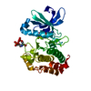
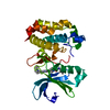
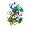
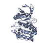
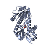
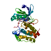
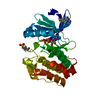
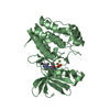
- PDB-6i2u: Aurora-A kinase domain in complex with Coenzyme A -
+
データを開く
IDまたはキーワード:
読み込み中...
-
基本情報
登録情報
データベース: PDB / ID: 6i2u
タイトル
Aurora-A kinase domain in complex with Coenzyme A
要素
Aurora kinase A
キーワード
TRANSFERASE / Aurora A Coenzyme A kinase covalent
機能・相同性
機能・相同性情報
Interaction between PHLDA1 and AURKA / regulation of centrosome cycle / axon hillock / spindle assembly involved in female meiosis I / cilium disassembly / spindle pole centrosome / histone H3S10 kinase activity / chromosome passenger complex / positive regulation of oocyte maturation / mitotic centrosome separation ...Interaction between PHLDA1 and AURKA / regulation of centrosome cycle / axon hillock / spindle assembly involved in female meiosis I / cilium disassembly / spindle pole centrosome / histone H3S10 kinase activity / chromosome passenger complex / positive regulation of oocyte maturation / mitotic centrosome separation / pronucleus / germinal vesicle / protein localization to centrosome / meiotic spindle / anterior/posterior axis specification / neuron projection extension / spindle organization / centrosome localization / positive regulation of mitochondrial fission / mitotic spindle pole / spindle midzone / SUMOylation of DNA replication proteins / negative regulation of protein binding / regulation of G2/M transition of mitotic cell cycle / positive regulation of mitotic nuclear division / protein serine/threonine/tyrosine kinase activity / positive regulation of mitotic cell cycle / TP53 Regulates Transcription of Genes Involved in G2 Cell Cycle Arrest / liver regeneration / molecular function activator activity / AURKA Activation by TPX2 / peptidyl-serine phosphorylation / regulation of signal transduction by p53 class mediator / mitotic spindle organization / regulation of cytokinesis / centriole / regulation of protein stability / response to wounding / APC/C:Cdh1 mediated degradation of Cdc20 and other APC/C:Cdh1 targeted proteins in late mitosis/early G1 / FBXL7 down-regulates AURKA during mitotic entry and in early mitosis / kinetochore / G2/M transition of mitotic cell cycle / spindle / spindle pole / mitotic spindle / protein autophosphorylation / Regulation of PLK1 Activity at G2/M Transition / mitotic cell cycle / positive regulation of proteasomal ubiquitin-dependent protein catabolic process / microtubule cytoskeleton / midbody / Regulation of TP53 Activity through Phosphorylation / basolateral plasma membrane / microtubule / protein phosphorylation / proteasome-mediated ubiquitin-dependent protein catabolic process / protein kinase activity / non-specific serine/threonine protein kinase / postsynaptic density / ciliary basal body / protein heterodimerization activity / negative regulation of gene expression / protein serine kinase activity / cell division / protein serine/threonine kinase activity / apoptotic process / ubiquitin protein ligase binding / centrosome / protein kinase binding / negative regulation of apoptotic process / perinuclear region of cytoplasm / glutamatergic synapse / nucleoplasm / ATP binding / nucleus / cytosol 類似検索 - 分子機能
Aurora kinase A / Aurora kinase / Phosphorylase Kinase; domain 1 / Phosphorylase Kinase; domain 1 / Transferase(Phosphotransferase) domain 1 / Transferase(Phosphotransferase); domain 1 / Serine/threonine-protein kinase, active site / Serine/Threonine protein kinases active-site signature. / Protein kinase domain / Serine/Threonine protein kinases, catalytic domain ...Aurora kinase A / Aurora kinase / Phosphorylase Kinase; domain 1 / Phosphorylase Kinase; domain 1 / Transferase(Phosphotransferase) domain 1 / Transferase(Phosphotransferase); domain 1 / Serine/threonine-protein kinase, active site / Serine/Threonine protein kinases active-site signature. / Protein kinase domain / Serine/Threonine protein kinases, catalytic domain / Protein kinase, ATP binding site / Protein kinases ATP-binding region signature. / Protein kinase domain profile. / Protein kinase domain / Protein kinase-like domain superfamily / 2-Layer Sandwich / Orthogonal Bundle / Mainly Alpha / Alpha Beta 類似検索 - ドメイン・相同性
COENZYME A / DI(HYDROXYETHYL)ETHER / Aurora kinase A 類似検索 - 構成要素
 ムービー
ムービー コントローラー


 データを開く
 基本情報
基本情報 要素
要素 キーワード
キーワード 機能・相同性情報
 Homo sapiens (ヒト)
 X線回折 /
  データ登録者
 英国, 1件
  引用
引用 構造の表示
構造の表示 ダウンロードとリンク
 その他のダウンロード
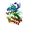

 PDBj
PDBj


 集合体
集合体


 分子量: 767.534 Da / 分子数: 1 / 由来タイプ: 合成 / 式: C21H36N7O16P3S
 分子量: 24.305 Da / 分子数: 4 / 由来タイプ: 合成 / 式: Mg
 分子量: 106.120 Da / 分子数: 3 / 由来タイプ: 合成 / 式: C4H10O3
 分子量: 18.015 Da / 分子数: 26 / 由来タイプ: 天然 / 式: H2O
 試料調製
試料調製 解析
解析