 Movie
Movie Controller

[English] 日本語
 Yorodumi
- PDB-4qd9: Crystal structure of Thioesterase PA1618 from Pseudomonas aerugin... -
+ Open data

- Basic information
| Entry | Database: PDB / ID: 4qd9 | ||||||
|---|---|---|---|---|---|---|---|
| Title | Crystal structure of Thioesterase PA1618 from Pseudomonas aeruginosa in complex with benzoyl-dO-CoA | ||||||
 Components Components | Thioesterase PA1618 | ||||||
 Keywords Keywords | HYDROLASE / Hotdog Fold / Thioesterase / Coenzyme A / acyl carrier protein / cytosol | ||||||
| Function / homology |  Function and homology information Function and homology information1,4-dihydroxy-2-naphthoyl-CoA thioesterase activity / Hydrolases; Acting on ester bonds; Thioester hydrolases / cytosol Similarity search - Function | ||||||
| Biological species |   Pseudomonas aeruginosa (bacteria) Pseudomonas aeruginosa (bacteria) | ||||||
| Method |  X-RAY DIFFRACTION / SYNCHROTRON / MOLECULAR REPLACEMENT / Resolution: 1.767 Å X-RAY DIFFRACTION / SYNCHROTRON / MOLECULAR REPLACEMENT / Resolution: 1.767 Å | ||||||
 Authors Authors | Ji, T. / Allen, K.N. / Dunaway-Mariano, D. | ||||||
 Citation Citation | Journal: To be Published Title: Design and Use of an Ester Analog of CoA to Trap the Michaelis Complex in a Thioesterase Authors: Latham, J.A. / Ji, T. / Matthews, K. / Allen, K.N. / Dunaway-Mariano, D. | ||||||
| History |
|
- Structure visualization
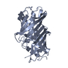
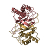
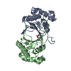
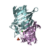
| Structure viewer | Molecule: MolmilJmol/JSmol |
|---|
- Downloads & links
-Download
| PDBx/mmCIF format | 4qd9.cif.gz | 180.6 KB | Display | PDBx/mmCIF format |
|---|---|---|---|---|
| PDB format | pdb4qd9.ent.gz | 146.4 KB | Display | PDB format |
| PDBx/mmJSON format | 4qd9.json.gz | Tree view | PDBx/mmJSON format | |
| Others |  Other downloads Other downloads |
-Validation report
| Arichive directory | https://data.pdbj.org/pub/pdb/validation_reports/qd/4qd9ftp://data.pdbj.org/pub/pdb/validation_reports/qd/4qd9 | HTTPS FTP |
|---|
-Related structure data


-Links
 PDBj
PDBj
- Assembly
| Deposited unit | 
| ||||||||
|---|---|---|---|---|---|---|---|---|---|
| 1 | 
| ||||||||
| 2 | 
| ||||||||
| 3 | 
| ||||||||
| Unit cell |
| ||||||||
| Components on special symmetry positions |
|
-Components
| #1: Protein | Mass: 16626.814 Da / Num. of mol.: 6 Source method: isolated from a genetically manipulated source Source: (gene. exp.) Pseudomonas aeruginosa (bacteria) / Strain: ATCC 15692 / PAO1 / 1C / PRS 101 / LMG 12228 / Gene: PA1618 / Plasmid: pET23-a / Production host: References: UniProt: Q9I3A4, Hydrolases; Acting on ester bonds; Thioester hydrolases #2: Chemical |   Mass: 775.595 Da / Num. of mol.: 2 / Source method: obtained synthetically / Formula: C28H39N7O15P2 Mass: 775.595 Da / Num. of mol.: 2 / Source method: obtained synthetically / Formula: C28H39N7O15P2#3: Water | ChemComp-HOH / |  Mass: 18.015 Da / Num. of mol.: 661 / Source method: isolated from a natural source / Formula: H2O Mass: 18.015 Da / Num. of mol.: 661 / Source method: isolated from a natural source / Formula: H2O |
|---|
-Experimental details
-Experiment
| Experiment | Method: X-RAY DIFFRACTION / Number of used crystals: 1 |
|---|
- Sample preparation
| Crystal | Density Matthews: 2.39 Å3/Da / Density % sol: 48.46 % |
|---|---|
| Crystal grow | Method: vapor diffusion, hanging drop / pH: 6.5 Details: 45% PEG400, 0.1M Bis Tris, 10mM beta-mercaptoethanol, pH 6.5, VAPOR DIFFUSION, HANGING DROP |
-Data collection
| Diffraction | Mean temperature: 100 K |
|---|---|
| Diffraction source | Source: SYNCHROTRON / Site: SSRL  / Beamline: BL12-2 / Wavelength: 0.98 Å / Beamline: BL12-2 / Wavelength: 0.98 Å |
| Detector | Type: PSI PILATUS 6M / Detector: PIXEL / Date: Mar 28, 2013 |
| Radiation | Monochromator: Si(111) / Protocol: SINGLE WAVELENGTH / Monochromatic (M) / Laue (L): M / Scattering type: x-ray |
| Radiation wavelength | Wavelength: 0.98 Å / Relative weight: 1 |
| Reflection | Resolution: 1.72→37.53 Å / Num. obs: 98312 / % possible obs: 97.85 % / Observed criterion σ(F): 1 / Observed criterion σ(I): 1 |
- Processing
| Software |
| ||||||||||||||||||||||||||||||||||||||||||||||||||||||||||||||||||||||||||||||||||||||||||||||||||
|---|---|---|---|---|---|---|---|---|---|---|---|---|---|---|---|---|---|---|---|---|---|---|---|---|---|---|---|---|---|---|---|---|---|---|---|---|---|---|---|---|---|---|---|---|---|---|---|---|---|---|---|---|---|---|---|---|---|---|---|---|---|---|---|---|---|---|---|---|---|---|---|---|---|---|---|---|---|---|---|---|---|---|---|---|---|---|---|---|---|---|---|---|---|---|---|---|---|---|---|
| Refinement | Method to determine structure: MOLECULAR REPLACEMENT / Resolution: 1.767→37.529 Å / SU ML: 0.2 / σ(F): 1.35 / Phase error: 24.84 / Stereochemistry target values: ML
| ||||||||||||||||||||||||||||||||||||||||||||||||||||||||||||||||||||||||||||||||||||||||||||||||||
| Solvent computation | Shrinkage radii: 0.9 Å / VDW probe radii: 1.11 Å / Solvent model: FLAT BULK SOLVENT MODEL | ||||||||||||||||||||||||||||||||||||||||||||||||||||||||||||||||||||||||||||||||||||||||||||||||||
| Refinement step | Cycle: LAST / Resolution: 1.767→37.529 Å
| ||||||||||||||||||||||||||||||||||||||||||||||||||||||||||||||||||||||||||||||||||||||||||||||||||
| Refine LS restraints |
| ||||||||||||||||||||||||||||||||||||||||||||||||||||||||||||||||||||||||||||||||||||||||||||||||||
| LS refinement shell |
|
