 Movie
Movie Controller

[English] 日本語
 Yorodumi
- PDB-3v8t: Crystal Structure of Interleukin-2 Inducible T-cell Kinase Itk Ca... -
+ Open data

- Basic information
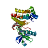
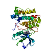
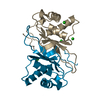
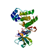
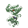
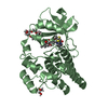
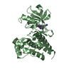
| Entry | Database: PDB / ID: 3v8t | ||||||
|---|---|---|---|---|---|---|---|
| Title | Crystal Structure of Interleukin-2 Inducible T-cell Kinase Itk Catalytic Domain with Thienopyrazolylindole Inhibitor 477 | ||||||
 Components Components | Tyrosine-protein kinase ITK/TSK | ||||||
 Keywords Keywords | TRANSFERASE/TRANSFERASE INHIBITOR / kinase / TRANSFERASE-TRANSFERASE INHIBITOR complex | ||||||
| Function / homology |  Function and homology information Function and homology informationNK T cell differentiation / gamma-delta T cell activation / Generation of second messenger molecules / cellular defense response / FCERI mediated Ca+2 mobilization / positive regulation of cytokine production / T cell activation / B cell receptor signaling pathway / non-specific protein-tyrosine kinase / non-membrane spanning protein tyrosine kinase activity ...NK T cell differentiation / gamma-delta T cell activation / Generation of second messenger molecules / cellular defense response / FCERI mediated Ca+2 mobilization / positive regulation of cytokine production / T cell activation / B cell receptor signaling pathway / non-specific protein-tyrosine kinase / non-membrane spanning protein tyrosine kinase activity / cell-cell junction / T cell receptor signaling pathway / adaptive immune response / intracellular signal transduction / signal transduction / zinc ion binding / ATP binding / nucleus / plasma membrane / cytosol Similarity search - Function | ||||||
| Biological species |  Homo sapiens (human) Homo sapiens (human) | ||||||
| Method |  X-RAY DIFFRACTION / SYNCHROTRON / MOLECULAR REPLACEMENT / Resolution: 2 Å X-RAY DIFFRACTION / SYNCHROTRON / MOLECULAR REPLACEMENT / Resolution: 2 Å | ||||||
 Authors Authors | McLean, L.R. / Zhang, Y. | ||||||
 Citation Citation | Journal: Bioorg.Med.Chem.Lett. / Year: 2012 Title: X-ray crystallographic structure-based design of selective thienopyrazole inhibitors for interleukin-2-inducible tyrosine kinase. Authors: McLean, L.R. / Zhang, Y. / Zaidi, N. / Bi, X. / Wang, R. / Dharanipragada, R. / Jurcak, J.G. / Gillespy, T.A. / Zhao, Z. / Musick, K.Y. / Choi, Y.M. / Barrague, M. / Peppard, J. / Smicker, M. ...Authors: McLean, L.R. / Zhang, Y. / Zaidi, N. / Bi, X. / Wang, R. / Dharanipragada, R. / Jurcak, J.G. / Gillespy, T.A. / Zhao, Z. / Musick, K.Y. / Choi, Y.M. / Barrague, M. / Peppard, J. / Smicker, M. / Duguid, M. / Parkar, A. / Fordham, J. / Kominos, D. | ||||||
| History |
|
- Structure visualization
| Structure viewer | Molecule: MolmilJmol/JSmol |
|---|
- Downloads & links
-Download
| PDBx/mmCIF format | 3v8t.cif.gz | 114.4 KB | Display | PDBx/mmCIF format |
|---|---|---|---|---|
| PDB format | pdb3v8t.ent.gz | 86.5 KB | Display | PDB format |
| PDBx/mmJSON format | 3v8t.json.gz | Tree view | PDBx/mmJSON format | |
| Others |  Other downloads Other downloads |
-Validation report
| Arichive directory | https://data.pdbj.org/pub/pdb/validation_reports/v8/3v8tftp://data.pdbj.org/pub/pdb/validation_reports/v8/3v8t | HTTPS FTP |
|---|
-Related structure data
| Related structure data |  3v5jC  3v5lC  3v8wC 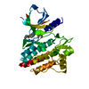 3vf8C 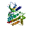 3vf9C 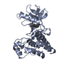 1sm2S C: citing same article ( S: Starting model for refinement |
|---|---|
| Similar structure data |


-Links
 PDBj
PDBj


- Assembly
| Deposited unit | 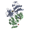
| ||||||||
|---|---|---|---|---|---|---|---|---|---|
| 1 | 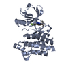
| ||||||||
| 2 | 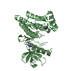
| ||||||||
| 3 |
| ||||||||
| Unit cell |
|
-Components
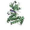
| #1: Protein | Mass: 30248.551 Da / Num. of mol.: 2 / Fragment: unp residues 357-620 Source method: isolated from a genetically manipulated source Source: (gene. exp.) Homo sapiens (human) / Gene: ITK, EMT, LYK / Plasmid: pFastBac1 / Production host:   Spodoptera frugiperda (fall armyworm) Spodoptera frugiperda (fall armyworm)References: UniProt: Q08881, non-specific protein-tyrosine kinase #2: Chemical | 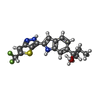  Mass: 375.435 Da / Num. of mol.: 2 / Source method: obtained synthetically / Formula: C19H19F2N3OS Mass: 375.435 Da / Num. of mol.: 2 / Source method: obtained synthetically / Formula: C19H19F2N3OS#3: Chemical | ChemComp-SO4 / |   Mass: 96.063 Da / Num. of mol.: 1 / Source method: obtained synthetically / Formula: SO4 Mass: 96.063 Da / Num. of mol.: 1 / Source method: obtained synthetically / Formula: SO4#4: Water | ChemComp-HOH / |  Mass: 18.015 Da / Num. of mol.: 276 / Source method: isolated from a natural source / Formula: H2O Mass: 18.015 Da / Num. of mol.: 276 / Source method: isolated from a natural source / Formula: H2O |
|---|
-Experimental details
-Experiment
| Experiment | Method: X-RAY DIFFRACTION / Number of used crystals: 1 |
|---|
- Sample preparation
| Crystal | Density Matthews: 2.73 Å3/Da / Density % sol: 54.87 % |
|---|---|
| Crystal grow | Temperature: 294 K / Method: vapor diffusion / pH: 6.2 Details: 0.8M Ammonium-sulfate, 0.1 M Na-citrate, 0.15 M Mg-acetate, 10 mM DTT, pH 6.2, VAPOR DIFFUSION, temperature 294K |
-Data collection
| Diffraction source | Source: SYNCHROTRON / Site: ESRF  / Beamline: ID14-1 / Wavelength: 0.934 Å / Beamline: ID14-1 / Wavelength: 0.934 Å | |||||||||||||||||||||||||||||||||||||||||||||||||||||||||||||||||||||||||||||
|---|---|---|---|---|---|---|---|---|---|---|---|---|---|---|---|---|---|---|---|---|---|---|---|---|---|---|---|---|---|---|---|---|---|---|---|---|---|---|---|---|---|---|---|---|---|---|---|---|---|---|---|---|---|---|---|---|---|---|---|---|---|---|---|---|---|---|---|---|---|---|---|---|---|---|---|---|---|---|
| Detector | Type: ADSC QUANTUM 4 / Detector: CCD / Date: Mar 8, 2005 | |||||||||||||||||||||||||||||||||||||||||||||||||||||||||||||||||||||||||||||
| Radiation | Protocol: SINGLE WAVELENGTH / Monochromatic (M) / Laue (L): M / Scattering type: x-ray | |||||||||||||||||||||||||||||||||||||||||||||||||||||||||||||||||||||||||||||
| Radiation wavelength | Wavelength: 0.934 Å / Relative weight: 1 | |||||||||||||||||||||||||||||||||||||||||||||||||||||||||||||||||||||||||||||
| Reflection | Resolution: 2→50 Å / Num. obs: 43469 / % possible obs: 98.4 % / Redundancy: 3.8 % / Rmerge(I) obs: 0.062 / Χ2: 1.472 / Net I/σ(I): 12.4 | |||||||||||||||||||||||||||||||||||||||||||||||||||||||||||||||||||||||||||||
| Reflection shell |
|
- Processing
| Software |
| ||||||||||||||||||||||||||||||||||||||||||||||||||||||||||||||||||||||||||||||||||||||||||||||||||||||||||||
|---|---|---|---|---|---|---|---|---|---|---|---|---|---|---|---|---|---|---|---|---|---|---|---|---|---|---|---|---|---|---|---|---|---|---|---|---|---|---|---|---|---|---|---|---|---|---|---|---|---|---|---|---|---|---|---|---|---|---|---|---|---|---|---|---|---|---|---|---|---|---|---|---|---|---|---|---|---|---|---|---|---|---|---|---|---|---|---|---|---|---|---|---|---|---|---|---|---|---|---|---|---|---|---|---|---|---|---|---|---|
| Refinement | Method to determine structure: MOLECULAR REPLACEMENT Starting model: pdb entry 1sm2 Resolution: 2→29.28 Å / Occupancy max: 1 / Occupancy min: 0.5 / SU R Cruickshank DPI: 0.169 / Cross valid method: THROUGHOUT / σ(F): 0
| ||||||||||||||||||||||||||||||||||||||||||||||||||||||||||||||||||||||||||||||||||||||||||||||||||||||||||||
| Displacement parameters | Biso max: 151.08 Å2 / Biso mean: 31.3413 Å2 / Biso min: 10.57 Å2
| ||||||||||||||||||||||||||||||||||||||||||||||||||||||||||||||||||||||||||||||||||||||||||||||||||||||||||||
| Refine analyze | Luzzati coordinate error obs: 0.274 Å | ||||||||||||||||||||||||||||||||||||||||||||||||||||||||||||||||||||||||||||||||||||||||||||||||||||||||||||
| Refinement step | Cycle: LAST / Resolution: 2→29.28 Å
| ||||||||||||||||||||||||||||||||||||||||||||||||||||||||||||||||||||||||||||||||||||||||||||||||||||||||||||
| Refine LS restraints |
| ||||||||||||||||||||||||||||||||||||||||||||||||||||||||||||||||||||||||||||||||||||||||||||||||||||||||||||
| LS refinement shell | Resolution: 2→2.05 Å / Total num. of bins used: 20
|
