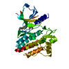
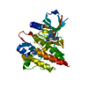
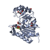
Entry Database : PDB / ID : 3vf9Title Crystal Structure of Spleen Tyrosine Kinase Syk Catalytic Domain with Thienopyrazolylindole Inhibitor 027 Tyrosine-protein kinase SYK Keywords / / Function / homology Function Domain/homology Component
/ / / / / / / / / / / / / / / / / / / / / / / / / / / / / / / / / / / / / / / / / / / / / / / / / / / / / / / / / / / / / / / / / / / / / / / / / / / / / / / / / / / / / / / / / / / / / / / / / / / / / / / / / / / / / / / / / / / / / / / / / / / / / / / / / / / / / / / / / / / / / / / / Biological species Homo sapiens (human)Method / / / Resolution : 2.3 Å Authors McLean, L.R. / Zhang, Y. Journal : Bioorg.Med.Chem.Lett. / Year : 2012Title : X-ray crystallographic structure-based design of selective thienopyrazole inhibitors for interleukin-2-inducible tyrosine kinase.Authors: McLean, L.R. / Zhang, Y. / Zaidi, N. / Bi, X. / Wang, R. / Dharanipragada, R. / Jurcak, J.G. / Gillespy, T.A. / Zhao, Z. / Musick, K.Y. / Choi, Y.M. / Barrague, M. / Peppard, J. / Smicker, M. ... Authors : McLean, L.R. / Zhang, Y. / Zaidi, N. / Bi, X. / Wang, R. / Dharanipragada, R. / Jurcak, J.G. / Gillespy, T.A. / Zhao, Z. / Musick, K.Y. / Choi, Y.M. / Barrague, M. / Peppard, J. / Smicker, M. / Duguid, M. / Parkar, A. / Fordham, J. / Kominos, D. History Deposition Jan 9, 2012 Deposition site / Processing site Revision 1.0 May 2, 2012 Provider / Type Revision 1.1 Nov 15, 2017 Group / Category / Item Revision 1.2 Feb 28, 2024 Group / Database references / Derived calculationsCategory chem_comp_atom / chem_comp_bond ... chem_comp_atom / chem_comp_bond / database_2 / struct_ref_seq_dif / struct_site Item _database_2.pdbx_DOI / _database_2.pdbx_database_accession ... _database_2.pdbx_DOI / _database_2.pdbx_database_accession / _struct_ref_seq_dif.details / _struct_site.pdbx_auth_asym_id / _struct_site.pdbx_auth_comp_id / _struct_site.pdbx_auth_seq_id
Show all Show less
 Movie
Movie Controller

 Yorodumi
 Open data
 Basic information
 Components
 Keywords
 Function and homology information
 Homo sapiens (human)
 X-RAY DIFFRACTION /
  Authors
 Citation
 Structure visualization
 Downloads & links
 Other downloads


 PDBj
PDBj


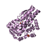
 Assembly

 Spodoptera frugiperda (fall armyworm)
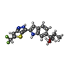
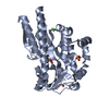
 Mass: 375.435 Da / Num. of mol.: 1 / Source method: obtained synthetically / Formula: C19H19F2N3OS
 Mass: 18.015 Da / Num. of mol.: 43 / Source method: isolated from a natural source / Formula: H2O
 Sample preparation
 Processing