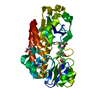
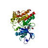
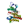
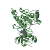
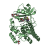
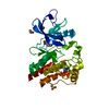
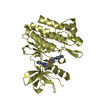
Entry Database : PDB / ID : 4xufTitle Crystal structure of the FLT3 kinase domain bound to the inhibitor quizartinib (AC220) Receptor-type tyrosine-protein kinase FLT3 Keywords / / / / / Function / homology Function Domain/homology Component
/ / / / / / / / / / / / / / / / / / / / / / / / / / / / / / / / / / / / / / / / / / / / / / / / / / / / / / / / / / / / / / / / / / / / / / / / / / / / / / / / / / / / / / / / / / / / / / / / / / / / / / / / / / / Biological species Homo sapiens (human)Method / / / Resolution : 3.2 Å Authors Zorn, J.A. / Wang, Q. / Fujimura, E. / Barros, T. / Kuriyan, J. Funding support Organization Grant number Country National Institutes of Health/National Cancer Institute (NIH/NCI) F32 CA177087-02 Cancer Research Institute Howard Hughes Medical Institute (HHMI)
Journal : Plos One / Year : 2015Title : Crystal Structure of the FLT3 Kinase Domain Bound to the Inhibitor Quizartinib (AC220).Authors : Zorn, J.A. / Wang, Q. / Fujimura, E. / Barros, T. / Kuriyan, J. History Deposition Jan 25, 2015 Deposition site / Processing site Revision 1.0 Apr 15, 2015 Provider / Type Revision 1.1 Apr 29, 2015 Group Revision 1.2 Aug 26, 2015 Group Revision 1.3 Sep 6, 2017 Group / Derived calculations / Category / pdbx_struct_oper_listItem / _pdbx_struct_oper_list.symmetry_operationRevision 1.4 Nov 20, 2019 Group / Category / Item Revision 1.5 Sep 27, 2023 Group Data collection / Database references ... Data collection / Database references / Refinement description / Structure summary Category chem_comp / chem_comp_atom ... chem_comp / chem_comp_atom / chem_comp_bond / database_2 / pdbx_initial_refinement_model / struct_ncs_dom_lim Item _chem_comp.pdbx_synonyms / _database_2.pdbx_DOI ... _chem_comp.pdbx_synonyms / _database_2.pdbx_DOI / _database_2.pdbx_database_accession / _struct_ncs_dom_lim.beg_auth_comp_id / _struct_ncs_dom_lim.beg_label_asym_id / _struct_ncs_dom_lim.beg_label_comp_id / _struct_ncs_dom_lim.beg_label_seq_id / _struct_ncs_dom_lim.end_auth_comp_id / _struct_ncs_dom_lim.end_label_asym_id / _struct_ncs_dom_lim.end_label_comp_id / _struct_ncs_dom_lim.end_label_seq_id
Show all Show less
 Movie
Movie Controller

 Yorodumi
 Open data
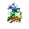
 Basic information
 Components
 Keywords
 Function and homology information
 Homo sapiens (human)
 X-RAY DIFFRACTION /
  Authors
 United States, 3items
  Citation
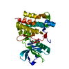
 Structure visualization
 Downloads & links
 Other downloads


 PDBj
PDBj


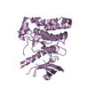
 Assembly
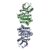


 unidentified baculovirus
 Mass: 560.667 Da / Num. of mol.: 2
 Mass: 18.015 Da / Num. of mol.: 2 / Source method: isolated from a natural source / Formula: H2O
 Sample preparation
 Processing