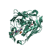
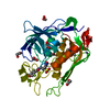
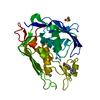
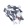
- PDB-4okg: LpxC from P.aeruginosa with the inhibitor 6-(benzimidazol-1-yl)-5... -
+
Open data
ID or keywords:
Loading...
-
Basic information
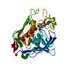
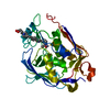
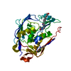
Entry
Database: PDB / ID: 4okg
Title
LpxC from P.aeruginosa with the inhibitor 6-(benzimidazol-1-yl)-5-[4-[2-[6-[(4-methylpiperazin-1-yl)methyl]-3-pyridyl]ethynyl]phenyl]pyridine-3-carbohydroxamic acid
UDP-3-O-acyl-N-acetylglucosamine deacetylase / UDP-3-O-acyl-N-acetylglucosamine deacetylase activity / lipid A biosynthetic process / membrane / metal ion binding / cytoplasm Similarity search - Function
Mass: 18.015 Da / Num. of mol.: 377 / Source method: isolated from a natural source / Formula: H2O
-
Experimental details
-
Experiment
Experiment
Method: X-RAY DIFFRACTION / Number of used crystals: 1
-
Sample preparation
Crystal
Density Matthews: 2.03 Å3/Da / Density % sol: 39.42 %
Crystal grow
Temperature: 293 K / Method: vapor diffusion, sitting drop / pH: 8.3 Details: Cocrystallization; protein in the following buffer: 20mM HEPES pH7.3, 150 mM NaCl, 2mM DTT, 10% Glycerol and concentrated to 11.3 mg/ml. Well solution contained: 2.2M ammonium sulfate, 0.1 M ...Details: Cocrystallization; protein in the following buffer: 20mM HEPES pH7.3, 150 mM NaCl, 2mM DTT, 10% Glycerol and concentrated to 11.3 mg/ml. Well solution contained: 2.2M ammonium sulfate, 0.1 M Tris pH 8.3, 4.1% PEG 400, 29 mM NaPO4 di-basic. Protein coexpressed with compound and well solution were combined 1:1 as a sitting drop. Crystals harvested approximately 21 days later., VAPOR DIFFUSION, SITTING DROP, temperature 293K
In the structure databanks used in Yorodumi, some data are registered as the other names, "COVID-19 virus" and "2019-nCoV". Here are the details of the virus and the list of structure data.
Jan 31, 2019. EMDB accession codes are about to change! (news from PDBe EMDB page)
EMDB accession codes are about to change! (news from PDBe EMDB page)
The allocation of 4 digits for EMDB accession codes will soon come to an end. Whilst these codes will remain in use, new EMDB accession codes will include an additional digit and will expand incrementally as the available range of codes is exhausted. The current 4-digit format prefixed with “EMD-” (i.e. EMD-XXXX) will advance to a 5-digit format (i.e. EMD-XXXXX), and so on. It is currently estimated that the 4-digit codes will be depleted around Spring 2019, at which point the 5-digit format will come into force.
The EM Navigator/Yorodumi systems omit the EMD- prefix.
Related info.:Q: What is EMD? / ID/Accession-code notation in Yorodumi/EM Navigator
Yorodumi is a browser for structure data from EMDB, PDB, SASBDB, etc.
This page is also the successor to EM Navigator detail page, and also detail information page/front-end page for Omokage search.
The word "yorodu" (or yorozu) is an old Japanese word meaning "ten thousand". "mi" (miru) is to see.
Related info.:EMDB / PDB / SASBDB / Comparison of 3 databanks / Yorodumi Search / Aug 31, 2016. New EM Navigator & Yorodumi / Yorodumi Papers / Jmol/JSmol / Function and homology information / Changes in new EM Navigator and Yorodumi
 Movie
Movie Controller

 Yorodumi
 Open data
 Basic information
 Components
 Keywords
 Function and homology information
 Pseudomonas aeruginosa (bacteria)
 X-RAY DIFFRACTION /
  Authors
 Citation
 Structure visualization
 Downloads & links
 Other downloads
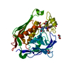


 PDBj
PDBj Assembly
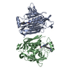


 Mass: 65.409 Da / Num. of mol.: 2 / Source method: obtained synthetically / Formula: Zn
 Mass: 543.618 Da / Num. of mol.: 2 / Source method: obtained synthetically / Formula: C32H29N7O2
 Mass: 18.015 Da / Num. of mol.: 377 / Source method: isolated from a natural source / Formula: H2O
 Sample preparation
 Processing