 Movie
Movie Controller

[English] 日本語
 Yorodumi
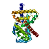
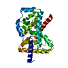
- PDB-4o42: Tandem chromodomains of human CHD1 in complex with influenza NS1 ... -
+ Open data

- Basic information
| Entry | Database: PDB / ID: 4o42 | ||||||
|---|---|---|---|---|---|---|---|
| Title | Tandem chromodomains of human CHD1 in complex with influenza NS1 C-terminal tail dimethylated at K229 | ||||||
 Components Components |
| ||||||
 Keywords Keywords | DNA BINDING PROTEIN/VIRAL PROTEIN / viral / chromodomain / Structural Genomics / Structural Genomics Consortium / SGC / DNA BINDING PROTEIN-VIRAL PROTEIN complex | ||||||
| Function / homology |  Function and homology information Function and homology informationsymbiont-mediated suppression of host mRNA processing / symbiont-mediated suppression of host PKR/eIFalpha signaling / : / nucleosome organization / host-mediated activation of viral transcription / histone H3K4me3 reader activity / ATP-dependent chromatin remodeler activity / nuclear chromosome / symbiont-mediated suppression of host cytoplasmic pattern recognition receptor signaling pathway via inhibition of RIG-I activity / protein serine/threonine kinase inhibitor activity ...symbiont-mediated suppression of host mRNA processing / symbiont-mediated suppression of host PKR/eIFalpha signaling / : / nucleosome organization / host-mediated activation of viral transcription / histone H3K4me3 reader activity / ATP-dependent chromatin remodeler activity / nuclear chromosome / symbiont-mediated suppression of host cytoplasmic pattern recognition receptor signaling pathway via inhibition of RIG-I activity / protein serine/threonine kinase inhibitor activity / Hydrolases; Acting on acid anhydrides; Acting on acid anhydrides to facilitate cellular and subcellular movement / histone binding / Estrogen-dependent gene expression / host cell cytoplasm / symbiont-mediated suppression of host type I interferon-mediated signaling pathway / chromatin remodeling / symbiont-mediated suppression of host gene expression / chromatin binding / chromatin / host cell nucleus / ATP hydrolysis activity / DNA binding / RNA binding / nucleoplasm / ATP binding / nucleus / cytoplasm Similarity search - Function | ||||||
| Biological species |  Homo sapiens (human) Homo sapiens (human)  Influenza A virus Influenza A virus | ||||||
| Method |  X-RAY DIFFRACTION / MOLECULAR REPLACEMENT / molecular replacement / Resolution: 1.87 Å X-RAY DIFFRACTION / MOLECULAR REPLACEMENT / molecular replacement / Resolution: 1.87 Å | ||||||
 Authors Authors | Qin, S. / Xu, C. / Tempel, W. / Arrowsmith, C.H. / Bountra, C. / Edwards, A.M. / Min, J. / Structural Genomics Consortium (SGC) | ||||||
 Citation Citation | Journal: Nat Commun / Year: 2014 Title: Structural basis for histone mimicry and hijacking of host proteins by influenza virus protein NS1. Authors: Qin, S. / Liu, Y. / Tempel, W. / Eram, M.S. / Bian, C. / Liu, K. / Senisterra, G. / Crombet, L. / Vedadi, M. / Min, J. | ||||||
| History |
|
- Structure visualization
| Structure viewer | Molecule: MolmilJmol/JSmol |
|---|
- Downloads & links
-Download
| PDBx/mmCIF format | 4o42.cif.gz | 90.6 KB | Display | PDBx/mmCIF format |
|---|---|---|---|---|
| PDB format | pdb4o42.ent.gz | 67.4 KB | Display | PDB format |
| PDBx/mmJSON format | 4o42.json.gz | Tree view | PDBx/mmJSON format | |
| Others |  Other downloads Other downloads |
-Validation report
| Arichive directory | https://data.pdbj.org/pub/pdb/validation_reports/o4/4o42ftp://data.pdbj.org/pub/pdb/validation_reports/o4/4o42 | HTTPS FTP |
|---|
-Related structure data
| Related structure data |  4nw2C  4o45C  4o46C 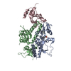 2b2wS S: Starting model for refinement C: citing same article ( |
|---|---|
| Similar structure data |


-Links
 PDBj
PDBj- Assembly
| Deposited unit | 
| ||||||||
|---|---|---|---|---|---|---|---|---|---|
| 1 |
| ||||||||
| Unit cell |
| ||||||||
| Components on special symmetry positions |
|
-Components
| #1: Protein | Mass: 22942.408 Da / Num. of mol.: 1 / Fragment: UNP residues 268-443 Source method: isolated from a genetically manipulated source Source: (gene. exp.) Homo sapiens (human) / Gene: CHD1 / Plasmid: pET28-MHL / Production host:  | ||||
|---|---|---|---|---|---|
| #2: Protein/peptide | Mass: 1819.269 Da / Num. of mol.: 1 / Fragment: UNP residues 216-230 / Source method: obtained synthetically / Details: synthetic peptide / Source: (synth.) Influenza A virus / References: UniProt: Q38SQ2 | ||||
| #3: Chemical | ChemComp-UNX /   Num. of mol.: 11 / Source method: obtained synthetically Num. of mol.: 11 / Source method: obtained synthetically#4: Chemical | ChemComp-GOL / |   Mass: 92.094 Da / Num. of mol.: 1 / Source method: obtained synthetically / Formula: C3H8O3 Mass: 92.094 Da / Num. of mol.: 1 / Source method: obtained synthetically / Formula: C3H8O3#5: Water | ChemComp-HOH / |  Mass: 18.015 Da / Num. of mol.: 96 / Source method: isolated from a natural source / Formula: H2O Mass: 18.015 Da / Num. of mol.: 96 / Source method: isolated from a natural source / Formula: H2O |
-Experimental details
-Experiment
| Experiment | Method: X-RAY DIFFRACTION / Number of used crystals: 1 |
|---|
- Sample preparation
| Crystal | Density Matthews: 2.8 Å3/Da / Density % sol: 56.2 % |
|---|---|
| Crystal grow | Temperature: 291 K / pH: 7 Details: 15% PEG3350, 0.1 succinic acid, pH 7, temperature 291K |
-Data collection
| Diffraction | Mean temperature: 100 K | ||||||||||||||||||
|---|---|---|---|---|---|---|---|---|---|---|---|---|---|---|---|---|---|---|---|
| Diffraction source | Source: ROTATING ANODE / Type: RIGAKU FR-E / Wavelength: 1.5418 Å | ||||||||||||||||||
| Detector | Type: RIGAKU SATURN A200 / Detector: CCD / Date: Jul 3, 2012 | ||||||||||||||||||
| Radiation | Protocol: SINGLE WAVELENGTH / Monochromatic (M) / Laue (L): M / Scattering type: x-ray | ||||||||||||||||||
| Radiation wavelength | Wavelength: 1.5418 Å / Relative weight: 1 | ||||||||||||||||||
| Reflection | Resolution: 1.87→46.06 Å / Num. obs: 20229 / % possible obs: 100 % / Redundancy: 7.1 % / Rmerge(I) obs: 0.087 / Net I/σ(I): 13.3 | ||||||||||||||||||
| Reflection shell | Rmerge(I) obs: 0.012 / Diffraction-ID: 1
|
-Phasing
| Phasing | Method: molecular replacement |
|---|
- Processing
| Software |
| |||||||||||||||||||||||||||||||||||||||||||||||||||||||||||||||||||||||||||
|---|---|---|---|---|---|---|---|---|---|---|---|---|---|---|---|---|---|---|---|---|---|---|---|---|---|---|---|---|---|---|---|---|---|---|---|---|---|---|---|---|---|---|---|---|---|---|---|---|---|---|---|---|---|---|---|---|---|---|---|---|---|---|---|---|---|---|---|---|---|---|---|---|---|---|---|---|
| Refinement | Method to determine structure: MOLECULAR REPLACEMENT Starting model: PDB ENTRY 2B2W Resolution: 1.87→46.06 Å / Cor.coef. Fo:Fc: 0.957 / Cor.coef. Fo:Fc free: 0.947 / WRfactor Rfree: 0.2131 / WRfactor Rwork: 0.1825 / Occupancy max: 1 / Occupancy min: 0.5 / FOM work R set: 0.8353 / SU B: 6.096 / SU ML: 0.094 / SU R Cruickshank DPI: 0.1249 / SU Rfree: 0.12 / Cross valid method: THROUGHOUT / σ(F): 0 / ESU R: 0.125 / ESU R Free: 0.12 / Stereochemistry target values: MAXIMUM LIKELIHOOD Details: coot was used for interactive model building. Model geometry was assessed with molprobity.
| |||||||||||||||||||||||||||||||||||||||||||||||||||||||||||||||||||||||||||
| Solvent computation | Ion probe radii: 0.8 Å / Shrinkage radii: 0.8 Å / VDW probe radii: 1.2 Å / Solvent model: MASK | |||||||||||||||||||||||||||||||||||||||||||||||||||||||||||||||||||||||||||
| Displacement parameters | Biso max: 130.25 Å2 / Biso mean: 41.1586 Å2 / Biso min: 17.48 Å2
| |||||||||||||||||||||||||||||||||||||||||||||||||||||||||||||||||||||||||||
| Refinement step | Cycle: LAST / Resolution: 1.87→46.06 Å
| |||||||||||||||||||||||||||||||||||||||||||||||||||||||||||||||||||||||||||
| Refine LS restraints |
| |||||||||||||||||||||||||||||||||||||||||||||||||||||||||||||||||||||||||||
| LS refinement shell | Resolution: 1.87→1.919 Å / Total num. of bins used: 20
| |||||||||||||||||||||||||||||||||||||||||||||||||||||||||||||||||||||||||||
| Refinement TLS params. | Method: refined / Origin x: -13.2516 Å / Origin y: -12.1436 Å / Origin z: 14.2216 Å
|
