 Movie
Movie Controller

[English] 日本語
 Yorodumi
- PDB-4nwo: Computationally Designed Two-Component Self-Assembling Tetrahedra... -
+ Open data

- Basic information
| Entry | Database: PDB / ID: 4nwo | ||||||
|---|---|---|---|---|---|---|---|
| Title | Computationally Designed Two-Component Self-Assembling Tetrahedral Cage T33-15 | ||||||
 Components Components |
| ||||||
 Keywords Keywords | PROTEIN BINDING / two-component / self-assembling / tetrahedron / designed protein cage / computational design / protein engineering / multimerization / nanomaterial / nanostructure / molybdenum cofactor biosynthesis protein / mog / chorismate mutase / isomerase | ||||||
| Function / homology |  Function and homology information Function and homology informationmolybdopterin cofactor biosynthetic process / molybdopterin adenylyltransferase activity / chorismate metabolic process / molybdopterin adenylyltransferase / chorismate mutase / chorismate mutase activity / Mo-molybdopterin cofactor biosynthetic process / aromatic amino acid biosynthetic process / amino acid biosynthetic process / ATP binding ...molybdopterin cofactor biosynthetic process / molybdopterin adenylyltransferase activity / chorismate metabolic process / molybdopterin adenylyltransferase / chorismate mutase / chorismate mutase activity / Mo-molybdopterin cofactor biosynthetic process / aromatic amino acid biosynthetic process / amino acid biosynthetic process / ATP binding / metal ion binding / cytoplasm / cytosol Similarity search - Function | ||||||
| Biological species |  Shewanella oneidensis (bacteria) Shewanella oneidensis (bacteria) Thermus thermophilus (bacteria) Thermus thermophilus (bacteria) | ||||||
| Method |  X-RAY DIFFRACTION / SYNCHROTRON / MOLECULAR REPLACEMENT / molecular replacement / Resolution: 2.8 Å X-RAY DIFFRACTION / SYNCHROTRON / MOLECULAR REPLACEMENT / molecular replacement / Resolution: 2.8 Å | ||||||
 Authors Authors | McNamara, D.E. / King, N.P. / Bale, J.B. / Sheffler, W. / Baker, D. / Yeates, T.O. | ||||||
 Citation Citation | Journal: Nature / Year: 2014 Title: Accurate design of co-assembling multi-component protein nanomaterials. Authors: King, N.P. / Bale, J.B. / Sheffler, W. / McNamara, D.E. / Gonen, S. / Gonen, T. / Yeates, T.O. / Baker, D. | ||||||
| History |
|
- Structure visualization
| Structure viewer | Molecule: MolmilJmol/JSmol |
|---|
- Downloads & links
-Download
| PDBx/mmCIF format | 4nwo.cif.gz | 117.3 KB | Display | PDBx/mmCIF format |
|---|---|---|---|---|
| PDB format | pdb4nwo.ent.gz | 92.3 KB | Display | PDB format |
| PDBx/mmJSON format | 4nwo.json.gz | Tree view | PDBx/mmJSON format | |
| Others |  Other downloads Other downloads |
-Validation report
| Arichive directory | https://data.pdbj.org/pub/pdb/validation_reports/nw/4nwoftp://data.pdbj.org/pub/pdb/validation_reports/nw/4nwo | HTTPS FTP |
|---|
-Related structure data
| Related structure data | 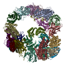 4nwnC 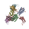 4nwpC 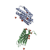 4nwqC  4nwrC  1ufyS 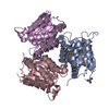 3k6aS C: citing same article ( S: Starting model for refinement |
|---|---|
| Similar structure data |


-Links
 PDBj
PDBj

- Assembly
| Deposited unit | 
| ||||||||
|---|---|---|---|---|---|---|---|---|---|
| 1 | x 12
| ||||||||
| Unit cell |
|
-Components
| #1: Protein | Mass: 19049.074 Da / Num. of mol.: 1 Mutation: Y20T, E21A, D30L, T31A, D34L, F110E, K135D, K137A, R140S, E141D, D144L, V168M Source method: isolated from a genetically manipulated source Source: (gene. exp.) Shewanella oneidensis (bacteria) / Gene: mogA, SO_0065 / Plasmid: pET29b / Production host: |
|---|---|
| #2: Protein | Mass: 14514.650 Da / Num. of mol.: 1 Mutation: E12N, E13S, E17T, A18S, H20I, Q21I, R24I, E25L, L28E, R109S Source method: isolated from a genetically manipulated source Source: (gene. exp.) Thermus thermophilus (bacteria) / Strain: HB8 / Gene: aroG, aroH / Plasmid: pET29b / Production host: References: UniProt: Q84FH6, UniProt: Q5SJY4*PLUS, chorismate mutase |
| #3: Chemical | ChemComp-CA /   Mass: 40.078 Da / Num. of mol.: 1 / Source method: obtained synthetically / Formula: Ca Mass: 40.078 Da / Num. of mol.: 1 / Source method: obtained synthetically / Formula: Ca |
| #4: Water | ChemComp-HOH /  Mass: 18.015 Da / Num. of mol.: 2 / Source method: isolated from a natural source / Formula: H2O Mass: 18.015 Da / Num. of mol.: 2 / Source method: isolated from a natural source / Formula: H2O |
-Experimental details
-Experiment
| Experiment | Method: X-RAY DIFFRACTION / Number of used crystals: 1 |
|---|
- Sample preparation
| Crystal | Density Matthews: 3.02 Å3/Da / Density % sol: 59.29 % |
|---|---|
| Crystal grow | Temperature: 298 K / pH: 6.5 Details: 100 mM sodium cacodylate pH 6.5, 200 mM calcium acetate, 28% (v/v) PEG 300, vapor diffusion, hanging drop, temperature 298K |
-Data collection
| Diffraction | Mean temperature: 100 K |
|---|---|
| Diffraction source | Source: SYNCHROTRON / Site: APS  / Beamline: 24-ID-C / Wavelength: 0.9792 / Beamline: 24-ID-C / Wavelength: 0.9792 |
| Detector | Type: DECTRIS PILATUS 6M / Detector: PIXEL / Date: Jun 9, 2013 |
| Radiation | Protocol: SINGLE WAVELENGTH / Monochromatic (M) / Laue (L): M / Scattering type: x-ray |
| Radiation wavelength | Wavelength: 0.9792 Å / Relative weight: 1 |
| Reflection | Resolution: 2.8→75.491 Å / Num. obs: 10783 / % possible obs: 99.9 % / Observed criterion σ(I): -3 / Redundancy: 13.59 % / Biso Wilson estimate: 79.22 Å2 / Rmerge(I) obs: 0.123 / Net I/σ(I): 19.43 |
| Reflection shell | Resolution: 2.8→2.87 Å / Redundancy: 14.34 % / Rmerge(I) obs: 1.863 / Mean I/σ(I) obs: 2.25 / % possible all: 100 |
-Phasing
| Phasing | Method: molecular replacement | |||||||||
|---|---|---|---|---|---|---|---|---|---|---|
| Phasing MR | Model details: Phaser MODE: MR_AUTO
|
- Processing
| Software |
| |||||||||||||||||||||||||||||||||||||||||||||||||||||||||||||||||||||||||||
|---|---|---|---|---|---|---|---|---|---|---|---|---|---|---|---|---|---|---|---|---|---|---|---|---|---|---|---|---|---|---|---|---|---|---|---|---|---|---|---|---|---|---|---|---|---|---|---|---|---|---|---|---|---|---|---|---|---|---|---|---|---|---|---|---|---|---|---|---|---|---|---|---|---|---|---|---|
| Refinement | Method to determine structure: MOLECULAR REPLACEMENT Starting model: 3K6A AND 1UFY Resolution: 2.8→75.49 Å / Occupancy max: 1 / Occupancy min: 1 / SU ML: 0.38 / σ(F): 1.34 / Phase error: 25.38 / Stereochemistry target values: ML
| |||||||||||||||||||||||||||||||||||||||||||||||||||||||||||||||||||||||||||
| Solvent computation | Shrinkage radii: 0.9 Å / VDW probe radii: 1.11 Å / Solvent model: FLAT BULK SOLVENT MODEL | |||||||||||||||||||||||||||||||||||||||||||||||||||||||||||||||||||||||||||
| Displacement parameters | Biso mean: 72.65 Å2 | |||||||||||||||||||||||||||||||||||||||||||||||||||||||||||||||||||||||||||
| Refinement step | Cycle: LAST / Resolution: 2.8→75.49 Å
| |||||||||||||||||||||||||||||||||||||||||||||||||||||||||||||||||||||||||||
| Refine LS restraints |
| |||||||||||||||||||||||||||||||||||||||||||||||||||||||||||||||||||||||||||
| LS refinement shell |
| |||||||||||||||||||||||||||||||||||||||||||||||||||||||||||||||||||||||||||
| Refinement TLS params. | Method: refined / Refine-ID: X-RAY DIFFRACTION
| |||||||||||||||||||||||||||||||||||||||||||||||||||||||||||||||||||||||||||
| Refinement TLS group |
|
