 Movie
Movie Controller

[English] 日本語
 Yorodumi
- PDB-4mva: 1.43 Angstrom Resolution Crystal Structure of Triosephosphate Iso... -
+ Open data

- Basic information
| Entry | Database: PDB / ID: 4mva | ||||||
|---|---|---|---|---|---|---|---|
| Title | 1.43 Angstrom Resolution Crystal Structure of Triosephosphate Isomerase (tpiA) from Escherichia coli in Complex with Acetyl Phosphate. | ||||||
 Components Components | Triosephosphate isomerase | ||||||
 Keywords Keywords | ISOMERASE / Structural Genomics / NIAID / National Institute of Allergy and Infectious Diseases / Center for Structural Genomics of Infectious Diseases / CSGID / TIM beta/alpha barrel / triose-phosphate isomerase activity | ||||||
| Function / homology | Aldolase class I / TIM Barrel / Alpha-Beta Barrel / Alpha Beta / DI(HYDROXYETHYL)ETHER / PHOSPHATE ION / ACETYLPHOSPHATE / :  Function and homology information Function and homology information | ||||||
| Biological species |  | ||||||
| Method |  X-RAY DIFFRACTION / SYNCHROTRON / MOLECULAR REPLACEMENT / Resolution: 1.43 Å X-RAY DIFFRACTION / SYNCHROTRON / MOLECULAR REPLACEMENT / Resolution: 1.43 Å | ||||||
 Authors Authors | Minasov, G. / Kuhn, M.L. / Dubrovska, I. / Winsor, J. / Shuvalova, L. / Grimshaw, S. / Kwon, K. / Anderson, W.F. / Center for Structural Genomics of Infectious Diseases (CSGID) | ||||||
 Citation Citation | Journal: Plos One / Year: 2014 Title: Structural, kinetic and proteomic characterization of acetyl phosphate-dependent bacterial protein acetylation. Authors: Kuhn, M.L. / Zemaitaitis, B. / Hu, L.I. / Sahu, A. / Sorensen, D. / Minasov, G. / Lima, B.P. / Scholle, M. / Mrksich, M. / Anderson, W.F. / Gibson, B.W. / Schilling, B. / Wolfe, A.J. | ||||||
| History |
|
- Structure visualization
| Structure viewer | Molecule: MolmilJmol/JSmol |
|---|
- Downloads & links
-Download
| PDBx/mmCIF format | 4mva.cif.gz | 239.2 KB | Display | PDBx/mmCIF format |
|---|---|---|---|---|
| PDB format | pdb4mva.ent.gz | 192.8 KB | Display | PDB format |
| PDBx/mmJSON format | 4mva.json.gz | Tree view | PDBx/mmJSON format | |
| Others |  Other downloads Other downloads |
-Validation report
| Arichive directory | https://data.pdbj.org/pub/pdb/validation_reports/mv/4mvaftp://data.pdbj.org/pub/pdb/validation_reports/mv/4mva | HTTPS FTP |
|---|
-Related structure data
| Related structure data |  4k6aC  4mvjC  1treS C: citing same article ( S: Starting model for refinement |
|---|---|
| Similar structure data | |
| Other databases |


-Links
 PDBj
PDBj- Assembly
| Deposited unit | 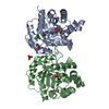
| ||||||||
|---|---|---|---|---|---|---|---|---|---|
| 1 |
| ||||||||
| Unit cell |
|
-Components
-Protein , 1 types, 2 molecules AB
| #1: Protein | Mass: 29747.729 Da / Num. of mol.: 2 / Fragment: Triosephosphate Isomerase (tpiA) Source method: isolated from a genetically manipulated source Source: (gene. exp.) |
|---|
-Non-polymers , 6 types, 844 molecules 


| #2: Chemical | ChemComp-CL /  Mass: 35.453 Da / Num. of mol.: 1 / Source method: obtained synthetically / Formula: Cl Mass: 35.453 Da / Num. of mol.: 1 / Source method: obtained synthetically / Formula: Cl | ||||||||
|---|---|---|---|---|---|---|---|---|---|
| #3: Chemical |  Mass: 140.032 Da / Num. of mol.: 2 / Source method: obtained synthetically / Formula: C2H5O5P Mass: 140.032 Da / Num. of mol.: 2 / Source method: obtained synthetically / Formula: C2H5O5P#4: Chemical | ChemComp-PO4 / |  Mass: 94.971 Da / Num. of mol.: 1 / Source method: obtained synthetically / Formula: PO4 Mass: 94.971 Da / Num. of mol.: 1 / Source method: obtained synthetically / Formula: PO4#5: Chemical |  Mass: 62.068 Da / Num. of mol.: 2 / Source method: obtained synthetically / Formula: C2H6O2 Mass: 62.068 Da / Num. of mol.: 2 / Source method: obtained synthetically / Formula: C2H6O2#6: Chemical | ChemComp-PEG / |  Mass: 106.120 Da / Num. of mol.: 1 / Source method: obtained synthetically / Formula: C4H10O3 Mass: 106.120 Da / Num. of mol.: 1 / Source method: obtained synthetically / Formula: C4H10O3#7: Water | ChemComp-HOH / | Mass: 18.015 Da / Num. of mol.: 837 / Source method: isolated from a natural source / Formula: H2O |
-Experimental details
-Experiment
| Experiment | Method: X-RAY DIFFRACTION / Number of used crystals: 1 |
|---|
- Sample preparation
| Crystal | Density Matthews: 1.99 Å3/Da / Density % sol: 38.04 % |
|---|---|
| Crystal grow | Temperature: 295 K / Method: vapor diffusion, sitting drop / pH: 6.5 Details: Protein: 7.4mg/mL, 0.15M Sodium cloride, 0.01M Tris-HCl pH 8.3; Screen: Classics II (F11), 0.2M Sodium chloride, 0.1M Bis-Tris pH 6.5, 25% (w/v) PEG 3350, 10mM ACP, VAPOR DIFFUSION, SITTING ...Details: Protein: 7.4mg/mL, 0.15M Sodium cloride, 0.01M Tris-HCl pH 8.3; Screen: Classics II (F11), 0.2M Sodium chloride, 0.1M Bis-Tris pH 6.5, 25% (w/v) PEG 3350, 10mM ACP, VAPOR DIFFUSION, SITTING DROP, temperature 295K |
-Data collection
| Diffraction | Mean temperature: 100 K |
|---|---|
| Diffraction source | Source: SYNCHROTRON / Site: APS  / Beamline: 21-ID-G / Wavelength: 0.97856 Å / Beamline: 21-ID-G / Wavelength: 0.97856 Å |
| Detector | Type: MARMOSAIC 300 mm CCD / Detector: CCD / Date: Mar 3, 2013 / Details: Beryllium lenses |
| Radiation | Monochromator: Diamond / Protocol: SINGLE WAVELENGTH / Monochromatic (M) / Laue (L): M / Scattering type: x-ray |
| Radiation wavelength | Wavelength: 0.97856 Å / Relative weight: 1 |
| Reflection | Resolution: 1.43→30 Å / Num. all: 88659 / Num. obs: 88659 / % possible obs: 99.9 % / Observed criterion σ(I): -3 / Redundancy: 6.1 % / Biso Wilson estimate: 16.2 Å2 / Rmerge(I) obs: 0.062 / Net I/σ(I): 22.7 |
| Reflection shell | Resolution: 1.43→1.45 Å / Redundancy: 6 % / Rmerge(I) obs: 0.534 / Mean I/σ(I) obs: 3.4 / Num. unique all: 4416 / % possible all: 100 |
- Processing
| Software |
| |||||||||||||||||||||||||||||||||||||||||||||||||||||||||||||||||||||||||||||||||||||||||||||||||||||||||||||||||||||||||||||
|---|---|---|---|---|---|---|---|---|---|---|---|---|---|---|---|---|---|---|---|---|---|---|---|---|---|---|---|---|---|---|---|---|---|---|---|---|---|---|---|---|---|---|---|---|---|---|---|---|---|---|---|---|---|---|---|---|---|---|---|---|---|---|---|---|---|---|---|---|---|---|---|---|---|---|---|---|---|---|---|---|---|---|---|---|---|---|---|---|---|---|---|---|---|---|---|---|---|---|---|---|---|---|---|---|---|---|---|---|---|---|---|---|---|---|---|---|---|---|---|---|---|---|---|---|---|---|
| Refinement | Method to determine structure: MOLECULAR REPLACEMENT Starting model: PDB ENTRY 1TRE Resolution: 1.43→29.24 Å / Cor.coef. Fo:Fc: 0.979 / Cor.coef. Fo:Fc free: 0.973 / SU B: 1.842 / SU ML: 0.037 Isotropic thermal model: Thermal Factors Isotropically Individually Refined Cross valid method: THROUGHOUT / ESU R: 0.057 / ESU R Free: 0.057 / Stereochemistry target values: MAXIMUM LIKELIHOOD / Details: HYDROGENS HAVE BEEN ADDED IN THE RIDING POSITIONS
| |||||||||||||||||||||||||||||||||||||||||||||||||||||||||||||||||||||||||||||||||||||||||||||||||||||||||||||||||||||||||||||
| Solvent computation | Ion probe radii: 0.8 Å / Shrinkage radii: 0.8 Å / VDW probe radii: 1.2 Å / Solvent model: MASK | |||||||||||||||||||||||||||||||||||||||||||||||||||||||||||||||||||||||||||||||||||||||||||||||||||||||||||||||||||||||||||||
| Displacement parameters | Biso mean: 19.023 Å2
| |||||||||||||||||||||||||||||||||||||||||||||||||||||||||||||||||||||||||||||||||||||||||||||||||||||||||||||||||||||||||||||
| Refinement step | Cycle: LAST / Resolution: 1.43→29.24 Å
| |||||||||||||||||||||||||||||||||||||||||||||||||||||||||||||||||||||||||||||||||||||||||||||||||||||||||||||||||||||||||||||
| Refine LS restraints |
| |||||||||||||||||||||||||||||||||||||||||||||||||||||||||||||||||||||||||||||||||||||||||||||||||||||||||||||||||||||||||||||
| LS refinement shell | Resolution: 1.43→1.467 Å / Total num. of bins used: 20
| |||||||||||||||||||||||||||||||||||||||||||||||||||||||||||||||||||||||||||||||||||||||||||||||||||||||||||||||||||||||||||||
| Refinement TLS params. | Method: refined / Refine-ID: X-RAY DIFFRACTION
| |||||||||||||||||||||||||||||||||||||||||||||||||||||||||||||||||||||||||||||||||||||||||||||||||||||||||||||||||||||||||||||
| Refinement TLS group |
|
