 Movie
Movie Controller


+ Open data

- Basic information
| Entry | Database: PDB / ID: 1b9b | ||||||
|---|---|---|---|---|---|---|---|
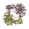
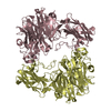
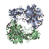
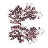
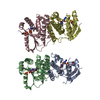
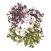
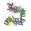
| Title | TRIOSEPHOSPHATE ISOMERASE OF THERMOTOGA MARITIMA | ||||||
 Components Components | PROTEIN (TRIOSEPHOSPHATE ISOMERASE) | ||||||
 Keywords Keywords | ISOMERASE / THERMOPHILIC / THERMOTOGA MARITIMA | ||||||
| Function / homology |  Function and homology information Function and homology informationphosphoglycerate kinase / phosphoglycerate kinase activity / triose-phosphate isomerase / triose-phosphate isomerase activity / gluconeogenesis / glycolytic process / ADP binding / fatty acid biosynthetic process / ATP binding / cytosol Similarity search - Function | ||||||
| Biological species |   Thermotoga maritima (bacteria) Thermotoga maritima (bacteria) | ||||||
| Method |  X-RAY DIFFRACTION / MOLECULAR REPLACEMENT / Resolution: 2.85 Å X-RAY DIFFRACTION / MOLECULAR REPLACEMENT / Resolution: 2.85 Å | ||||||
 Authors Authors | Maes, D. / Wierenga, R.K. | ||||||
 Citation Citation | Journal: Proteins / Year: 1999 Title: The crystal structure of triosephosphate isomerase (TIM) from Thermotoga maritima: a comparative thermostability structural analysis of ten different TIM structures. Authors: Maes, D. / Zeelen, J.P. / Thanki, N. / Beaucamp, N. / Alvarez, M. / Thi, M.H. / Backmann, J. / Martial, J.A. / Wyns, L. / Jaenicke, R. / Wierenga, R.K. | ||||||
| History |
|
- Structure visualization
| Structure viewer | Molecule: MolmilJmol/JSmol |
|---|
- Downloads & links
-Download
| PDBx/mmCIF format | 1b9b.cif.gz | 110 KB | Display | PDBx/mmCIF format |
|---|---|---|---|---|
| PDB format | pdb1b9b.ent.gz | 86.4 KB | Display | PDB format |
| PDBx/mmJSON format | 1b9b.json.gz | Tree view | PDBx/mmJSON format | |
| Others |  Other downloads Other downloads |
-Validation report
| Arichive directory | https://data.pdbj.org/pub/pdb/validation_reports/b9/1b9bftp://data.pdbj.org/pub/pdb/validation_reports/b9/1b9b | HTTPS FTP |
|---|
-Related structure data
| Related structure data | 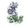 1bimS S: Starting model for refinement |
|---|---|
| Similar structure data |

-Links
 PDBj
PDBj
- Assembly
| Deposited unit | 
| ||||||||||
|---|---|---|---|---|---|---|---|---|---|---|---|
| 1 | 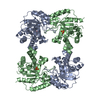
| ||||||||||
| 2 |
| ||||||||||
| Unit cell |
| ||||||||||
| Details | THIS TIM FROM A HYPERTHERMOPHILIC ORGANISM EXISTS IN VIVO AS A PGK-TIM FUSION PROTEIN. THE RECOMBINANT SEPARATE TIM-ENZYME IS ALSO HYPERTHERMOPHILIC. IT IS A TETRAMER. THE TETRAMER IS FORMED BY APPLYING THE CRYSTALLOGRAPHIC TWOFOLD AXIS ON THE ASYMETRIC UNIT (A DIMER). THE MAIN INTERDIMERIC CONTACTS ARE 2 DISULFIDE BONDS. |
-Components
| #1: Protein | Mass: 28554.045 Da / Num. of mol.: 2 Source method: isolated from a genetically manipulated source Details: A SULFATE MOLECULE IS OBSERVED IN THE ACTIVE SITE OF BOTH SUBUNITS Source: (gene. exp.) Thermotoga maritima (bacteria) / Production host: #2: Chemical |   Mass: 96.063 Da / Num. of mol.: 2 / Source method: obtained synthetically / Formula: SO4 Mass: 96.063 Da / Num. of mol.: 2 / Source method: obtained synthetically / Formula: SO4#3: Water | ChemComp-HOH / |  Mass: 18.015 Da / Num. of mol.: 52 / Source method: isolated from a natural source / Formula: H2O Mass: 18.015 Da / Num. of mol.: 52 / Source method: isolated from a natural source / Formula: H2OHas protein modification | Y | |
|---|
-Experimental details
-Experiment
| Experiment | Method: X-RAY DIFFRACTION / Number of used crystals: 1 |
|---|
- Sample preparation
| Crystal | Density Matthews: 4.16 Å3/Da / Density % sol: 70 % | ||||||||||||||||||||||||||||||||||||||||||||||||
|---|---|---|---|---|---|---|---|---|---|---|---|---|---|---|---|---|---|---|---|---|---|---|---|---|---|---|---|---|---|---|---|---|---|---|---|---|---|---|---|---|---|---|---|---|---|---|---|---|---|
| Crystal grow | pH: 7 / Details: pH 7.0 | ||||||||||||||||||||||||||||||||||||||||||||||||
| Crystal | *PLUS | ||||||||||||||||||||||||||||||||||||||||||||||||
| Crystal grow | *PLUS Temperature: 20 ℃ / pH: 8 / Method: vapor diffusion, hanging drop | ||||||||||||||||||||||||||||||||||||||||||||||||
| Components of the solutions | *PLUS
|
-Data collection
| Diffraction | Mean temperature: 298 K |
|---|---|
| Diffraction source | Source: ROTATING ANODE / Type: ENRAF-NONIUS FR571 / Wavelength: 1.5418 |
| Detector | Type: MARRESEARCH / Detector: IMAGE PLATE / Date: Nov 15, 1996 / Details: DOUBLE FOCUSSING MIRRORS |
| Radiation | Protocol: SINGLE WAVELENGTH / Monochromatic (M) / Laue (L): M / Scattering type: x-ray |
| Radiation wavelength | Wavelength: 1.5418 Å / Relative weight: 1 |
| Reflection | Resolution: 2.85→20 Å / Num. all: 22000 / Num. obs: 22000 / % possible obs: 97.8 % / Redundancy: 3.1 % / Biso Wilson estimate: 38.7 Å2 / Rmerge(I) obs: 0.086 / Rsym value: 0.07 / Net I/σ(I): 9 |
| Reflection shell | Resolution: 2.85→2.9 Å / Redundancy: 2.7 % / Rmerge(I) obs: 0.299 / Mean I/σ(I) obs: 6.7 / Rsym value: 0.247 / % possible all: 90 |
| Reflection | *PLUS Num. measured all: 88489 |
| Reflection shell | *PLUS % possible obs: 90 % |
- Processing
| Software |
| ||||||||||||||||||||||||||||||||||||||||||||||||||||||||||||
|---|---|---|---|---|---|---|---|---|---|---|---|---|---|---|---|---|---|---|---|---|---|---|---|---|---|---|---|---|---|---|---|---|---|---|---|---|---|---|---|---|---|---|---|---|---|---|---|---|---|---|---|---|---|---|---|---|---|---|---|---|---|
| Refinement | Method to determine structure: MOLECULAR REPLACEMENT Starting model: BACILLUS STEAROTHERMOPHILUS TIM (PDB ENTRY 1BIM) Resolution: 2.85→20 Å / Cross valid method: THROUGHOUT / σ(F): 0
| ||||||||||||||||||||||||||||||||||||||||||||||||||||||||||||
| Solvent computation | Solvent model: BULK SOLVENT MODEL / Bsol: 53 Å2 / ksol: 0.289 e/Å3 | ||||||||||||||||||||||||||||||||||||||||||||||||||||||||||||
| Refinement step | Cycle: LAST / Resolution: 2.85→20 Å
| ||||||||||||||||||||||||||||||||||||||||||||||||||||||||||||
| Refine LS restraints |
| ||||||||||||||||||||||||||||||||||||||||||||||||||||||||||||
| LS refinement shell | Resolution: 2.85→2.9 Å / Total num. of bins used: 21
| ||||||||||||||||||||||||||||||||||||||||||||||||||||||||||||
| Xplor file |
| ||||||||||||||||||||||||||||||||||||||||||||||||||||||||||||
| Software | *PLUS Name: CNS / Version: 0.4 / Classification: refinement | ||||||||||||||||||||||||||||||||||||||||||||||||||||||||||||
| Refinement | *PLUS Lowest resolution: 20 Å / σ(F): 0 / % reflection Rfree: 5 % / Rfactor obs: 0.211 | ||||||||||||||||||||||||||||||||||||||||||||||||||||||||||||
| Solvent computation | *PLUS | ||||||||||||||||||||||||||||||||||||||||||||||||||||||||||||
| Displacement parameters | *PLUS | ||||||||||||||||||||||||||||||||||||||||||||||||||||||||||||
| Refine LS restraints | *PLUS
| ||||||||||||||||||||||||||||||||||||||||||||||||||||||||||||
| LS refinement shell | *PLUS Rfactor Rfree: 0.33 / % reflection Rfree: 5 % / Rfactor Rwork: 0.36 |
