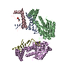
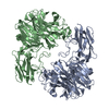
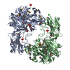
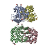
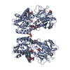
THE DIMER IN THIS ENTRY IS THE HEAD TO HEAD (HH)-DIMER THAT HAS SUBSEQUENTLY BEEN IDENTIFIED AS THE PHYSIOLOGICALLY ACTIVE DIMER BY SITE-DIRECTED MUTAGENESIS STUDIES (QIU, Y., OGAWA, H., ZHANG, X., MISONO, K.S. J.BIOL.CHEM. 279:6115-23, 2004) AND THE DIMER IN SOLUTION BY ELECTRON MICROSCOPY COUPLED WITH SINGLE PARTICLE RECONSTRUCTION (SUBMITTED FOR PUBLICATION). THE CC DIMER (biolomecule 1) WAS CHOSEN by author FOR A SLIGHTLY LARGER BURIED SURFACE AREA AND MORE CARBOXYL TERMINAL STRUCTURE ASSIGNED.
-
Components
-
Protein , 1 types, 2 molecules AC
#1: Protein
ATRIALNATRIURETICPEPTIDERECEPTORA
Mass: 48434.828 Da / Num. of mol.: 2 / Fragment: HORMONE BINDING DOMAIN Source method: isolated from a genetically manipulated source Source: (gene. exp.) Rattus norvegicus (Norway rat) / Plasmid: PCDNA3-NPR / Production host: Chlorocebus aethiops (grivet monkey) / References: UniProt: P18910, guanylate cyclase
In the structure databanks used in Yorodumi, some data are registered as the other names, "COVID-19 virus" and "2019-nCoV". Here are the details of the virus and the list of structure data.
Jan 31, 2019. EMDB accession codes are about to change! (news from PDBe EMDB page)
EMDB accession codes are about to change! (news from PDBe EMDB page)
The allocation of 4 digits for EMDB accession codes will soon come to an end. Whilst these codes will remain in use, new EMDB accession codes will include an additional digit and will expand incrementally as the available range of codes is exhausted. The current 4-digit format prefixed with “EMD-” (i.e. EMD-XXXX) will advance to a 5-digit format (i.e. EMD-XXXXX), and so on. It is currently estimated that the 4-digit codes will be depleted around Spring 2019, at which point the 5-digit format will come into force.
The EM Navigator/Yorodumi systems omit the EMD- prefix.
Related info.:Q: What is EMD? / ID/Accession-code notation in Yorodumi/EM Navigator
Yorodumi is a browser for structure data from EMDB, PDB, SASBDB, etc.
This page is also the successor to EM Navigator detail page, and also detail information page/front-end page for Omokage search.
The word "yorodu" (or yorozu) is an old Japanese word meaning "ten thousand". "mi" (miru) is to see.
Related info.:EMDB / PDB / SASBDB / Comparison of 3 databanks / Yorodumi Search / Aug 31, 2016. New EM Navigator & Yorodumi / Yorodumi Papers / Jmol/JSmol / Function and homology information / Changes in new EM Navigator and Yorodumi
 Movie
Movie Controller

 Yorodumi
 Open data
 Basic information
 Components
 Keywords
 Function and homology information
 X-RAY DIFFRACTION /
  Authors
 Citation
 Structure visualization
 Downloads & links
 Other downloads
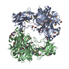


 PDBj
PDBj


 Assembly

 Type: D-saccharide, beta linking / Mass: 221.208 Da / Num. of mol.: 1


 Mass: 96.063 Da / Num. of mol.: 9 / Source method: obtained synthetically / Formula: SO4
 Mass: 35.453 Da / Num. of mol.: 2 / Source method: obtained synthetically / Formula: Cl
 Sample preparation
 / Beamline: 19-ID / Wavelength: 1
  Processing