[English] 日本語
 Yorodumi
- PDB-4msd: Crystal structure of Schizosaccharomyces pombe AMSH-like protein ... -
+ Open data
- Basic information
| Entry | Database: PDB / ID: 4msd | ||||||
|---|---|---|---|---|---|---|---|
| Title | Crystal structure of Schizosaccharomyces pombe AMSH-like protein SST2 T319I mutant | ||||||
 Components Components | AMSH-like protease sst2 | ||||||
 Keywords Keywords | HYDROLASE / Helix-beta-helix sandwich / ubiquitin / deubiquitination / zinc metalloprotease / AMSH / lysine 63-linked polyubiquitin / cytosol | ||||||
| Function / homology |  Function and homology information Function and homology informationMetalloprotease DUBs / cytoplasm to vacuole targeting by the NVT pathway / late endosome to vacuole transport via multivesicular body sorting pathway / protein transport to vacuole involved in ubiquitin-dependent protein catabolic process via the multivesicular body sorting pathway / Hydrolases; Acting on peptide bonds (peptidases); Omega peptidases / late endosome to vacuole transport / protein K63-linked deubiquitination / metal-dependent deubiquitinase activity / K63-linked polyubiquitin modification-dependent protein binding / K63-linked deubiquitinase activity ...Metalloprotease DUBs / cytoplasm to vacuole targeting by the NVT pathway / late endosome to vacuole transport via multivesicular body sorting pathway / protein transport to vacuole involved in ubiquitin-dependent protein catabolic process via the multivesicular body sorting pathway / Hydrolases; Acting on peptide bonds (peptidases); Omega peptidases / late endosome to vacuole transport / protein K63-linked deubiquitination / metal-dependent deubiquitinase activity / K63-linked polyubiquitin modification-dependent protein binding / K63-linked deubiquitinase activity / cell division site / ubiquitin binding / endosome / zinc ion binding / membrane / cytoplasm Similarity search - Function | ||||||
| Biological species |  | ||||||
| Method |  X-RAY DIFFRACTION / X-RAY DIFFRACTION /  SYNCHROTRON / SYNCHROTRON /  MOLECULAR REPLACEMENT / Resolution: 1.9 Å MOLECULAR REPLACEMENT / Resolution: 1.9 Å | ||||||
 Authors Authors | Shrestha, R.K. / Ronau, J.A. / Das, C. | ||||||
 Citation Citation |  Journal: Biochemistry / Year: 2014 Journal: Biochemistry / Year: 2014Title: Insights into the Mechanism of Deubiquitination by JAMM Deubiquitinases from Cocrystal Structures of the Enzyme with the Substrate and Product. Authors: Shrestha, R.K. / Ronau, J.A. / Davies, C.W. / Guenette, R.G. / Strieter, E.R. / Paul, L.N. / Das, C. | ||||||
| History |
|
- Structure visualization
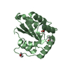
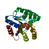
| Structure viewer | Molecule:  Molmil Molmil Jmol/JSmol Jmol/JSmol |
|---|
- Downloads & links
- Download
| PDBx/mmCIF format |  4msd.cif.gz 4msd.cif.gz | 93.2 KB | Display |  PDBx/mmCIF format PDBx/mmCIF format |
|---|---|---|---|---|
| PDB format |  pdb4msd.ent.gz pdb4msd.ent.gz | 69.8 KB | Display |  PDB format PDB format |
| PDBx/mmJSON format |  4msd.json.gz 4msd.json.gz | Tree view |  PDBx/mmJSON format PDBx/mmJSON format | |
| Others |  Other downloads Other downloads |
-Validation report
| Summary document |  4msd_validation.pdf.gz 4msd_validation.pdf.gz | 467.5 KB | Display |  wwPDB validaton report wwPDB validaton report |
|---|---|---|---|---|
| Full document |  4msd_full_validation.pdf.gz 4msd_full_validation.pdf.gz | 469.1 KB | Display | |
| Data in XML |  4msd_validation.xml.gz 4msd_validation.xml.gz | 17.6 KB | Display | |
| Data in CIF |  4msd_validation.cif.gz 4msd_validation.cif.gz | 24.3 KB | Display | |
| Arichive directory |  https://data.pdbj.org/pub/pdb/validation_reports/ms/4msd https://data.pdbj.org/pub/pdb/validation_reports/ms/4msd ftp://data.pdbj.org/pub/pdb/validation_reports/ms/4msd ftp://data.pdbj.org/pub/pdb/validation_reports/ms/4msd | HTTPS FTP |
-Related structure data
| Related structure data |  4jxeSC  4k1rC  4ms7C  4msjC  4msmC 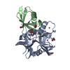 4msqC 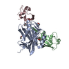 4nqlC 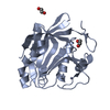 4pqtC S: Starting model for refinement C: citing same article ( |
|---|---|
| Similar structure data |
- Links
Links
- Assembly
| Deposited unit | 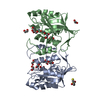
| ||||||||
|---|---|---|---|---|---|---|---|---|---|
| 1 | 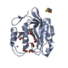
| ||||||||
| 2 | 
| ||||||||
| Unit cell |
|
- Components
| #1: Protein | Mass: 21993.787 Da / Num. of mol.: 2 / Fragment: catalytic domain, UNP residues 245-435 / Mutation: T319I Source method: isolated from a genetically manipulated source Source: (gene. exp.)  Strain: 972/ATCC 24843 / Gene: sst2, SPAC19B12.10 / Plasmid: pGEX-6P-1 / Production host:  References: UniProt: Q9P371, Hydrolases; Acting on peptide bonds (peptidases); Omega peptidases #2: Chemical | ChemComp-ZN / #3: Chemical | ChemComp-EDO / #4: Chemical | ChemComp-DTT / | #5: Water | ChemComp-HOH / | |
|---|
-Experimental details
-Experiment
| Experiment | Method:  X-RAY DIFFRACTION / Number of used crystals: 1 X-RAY DIFFRACTION / Number of used crystals: 1 |
|---|
- Sample preparation
| Crystal | Density Matthews: 2.91 Å3/Da / Density % sol: 57.75 % |
|---|---|
| Crystal grow | Temperature: 293 K / Method: vapor diffusion, sitting drop / pH: 7.6 Details: 0.03 M citric acid, 0.07 M bis-tris propane, pH 7.6, 20% PEG 3350, benzamidine hydrochloride (additive), VAPOR DIFFUSION, SITTING DROP, temperature 293K |
-Data collection
| Diffraction | Mean temperature: 100 K |
|---|---|
| Diffraction source | Source:  SYNCHROTRON / Site: SYNCHROTRON / Site:  APS APS  / Beamline: 23-ID-B / Wavelength: 1.033 Å / Beamline: 23-ID-B / Wavelength: 1.033 Å |
| Detector | Type: MAR scanner 300 mm plate / Detector: IMAGE PLATE / Date: Jul 7, 2013 |
| Radiation | Monochromator: Si 111 Channel / Protocol: SINGLE WAVELENGTH / Monochromatic (M) / Laue (L): M / Scattering type: x-ray |
| Radiation wavelength | Wavelength: 1.033 Å / Relative weight: 1 |
| Reflection | Resolution: 1.9→50 Å / Num. all: 39816 / Num. obs: 39816 / % possible obs: 100 % / Observed criterion σ(F): 2.5 / Observed criterion σ(I): 2.5 / Redundancy: 3.8 % / Rmerge(I) obs: 0.07 / Rsym value: 0.07 / Net I/σ(I): 18.5 |
| Reflection shell | Resolution: 1.9→1.93 Å / Redundancy: 3.8 % / Rmerge(I) obs: 0.565 / Mean I/σ(I) obs: 2.5 / Num. unique all: 1996 / Rsym value: 0.565 / % possible all: 100 |
- Processing
| Software |
| |||||||||||||||||||||||||||||||||||||||||||||||||||||||||||||||||||||||||||||||||||||||||||||||||||||||||
|---|---|---|---|---|---|---|---|---|---|---|---|---|---|---|---|---|---|---|---|---|---|---|---|---|---|---|---|---|---|---|---|---|---|---|---|---|---|---|---|---|---|---|---|---|---|---|---|---|---|---|---|---|---|---|---|---|---|---|---|---|---|---|---|---|---|---|---|---|---|---|---|---|---|---|---|---|---|---|---|---|---|---|---|---|---|---|---|---|---|---|---|---|---|---|---|---|---|---|---|---|---|---|---|---|---|---|
| Refinement | Method to determine structure:  MOLECULAR REPLACEMENT MOLECULAR REPLACEMENTStarting model: PDB Entry 4JXE Resolution: 1.9→33.612 Å / SU ML: 0.22 / Cross valid method: THROUGHOUT / σ(F): 1.38 / Phase error: 23.02 / Stereochemistry target values: ML
| |||||||||||||||||||||||||||||||||||||||||||||||||||||||||||||||||||||||||||||||||||||||||||||||||||||||||
| Solvent computation | Shrinkage radii: 0.9 Å / VDW probe radii: 1.11 Å / Solvent model: FLAT BULK SOLVENT MODEL | |||||||||||||||||||||||||||||||||||||||||||||||||||||||||||||||||||||||||||||||||||||||||||||||||||||||||
| Refinement step | Cycle: LAST / Resolution: 1.9→33.612 Å
| |||||||||||||||||||||||||||||||||||||||||||||||||||||||||||||||||||||||||||||||||||||||||||||||||||||||||
| Refine LS restraints |
| |||||||||||||||||||||||||||||||||||||||||||||||||||||||||||||||||||||||||||||||||||||||||||||||||||||||||
| LS refinement shell |
|
 Movie
Movie Controller






 PDBj
PDBj


