 Movie
Movie Controller

[English] 日本語
 Yorodumi
- PDB-4lz9: Structure of MATE multidrug transporter DinF-BH in complex with R6G -
+ Open data

- Basic information
| Entry | Database: PDB / ID: 4lz9 | ||||||
|---|---|---|---|---|---|---|---|
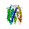
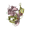
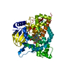
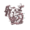
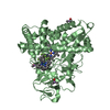
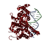
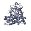
| Title | Structure of MATE multidrug transporter DinF-BH in complex with R6G | ||||||
 Components Components | BH2163 protein | ||||||
 Keywords Keywords | TRANSPORT PROTEIN / multidrug transporter | ||||||
| Function / homology |  Function and homology information Function and homology informationantiporter activity / xenobiotic transmembrane transporter activity / response to antibiotic / plasma membrane Similarity search - Function | ||||||
| Biological species |  Bacillus halodurans (bacteria) Bacillus halodurans (bacteria) | ||||||
| Method |  X-RAY DIFFRACTION / SYNCHROTRON / AB INITIO PHASING / Resolution: 3.7 Å X-RAY DIFFRACTION / SYNCHROTRON / AB INITIO PHASING / Resolution: 3.7 Å | ||||||
 Authors Authors | Lu, M. / Radchenko, M. / Symersky, J. / Nie, R. / Guo, Y. | ||||||
 Citation Citation | Journal: Nat.Struct.Mol.Biol. / Year: 2013 Title: Structural insights into H(+)-coupled multidrug extrusion by a MATE transporter Authors: Lu, M. / Radchenko, M. / Symersky, J. / Nie, R. / Guo, Y. | ||||||
| History |
|
- Structure visualization
| Structure viewer | Molecule: MolmilJmol/JSmol |
|---|
- Downloads & links
-Download
| PDBx/mmCIF format | 4lz9.cif.gz | 29.5 KB | Display | PDBx/mmCIF format |
|---|---|---|---|---|
| PDB format | pdb4lz9.ent.gz | 15 KB | Display | PDB format |
| PDBx/mmJSON format | 4lz9.json.gz | Tree view | PDBx/mmJSON format | |
| Others |  Other downloads Other downloads |
-Validation report
| Arichive directory | https://data.pdbj.org/pub/pdb/validation_reports/lz/4lz9ftp://data.pdbj.org/pub/pdb/validation_reports/lz/4lz9 | HTTPS FTP |
|---|
-Related structure data


-Links
 PDBj
PDBj
- Assembly
| Deposited unit | 
| ||||||||
|---|---|---|---|---|---|---|---|---|---|
| 1 |
| ||||||||
| Unit cell |
|
-Components
| #1: Protein | Mass: 48615.172 Da / Num. of mol.: 1 / Fragment: UNP residues 3-448 Source method: isolated from a genetically manipulated source Source: (gene. exp.) Bacillus halodurans (bacteria) / Strain: C-125 / Gene: BH2163 / Production host: |
|---|---|
| #2: Chemical | ChemComp-RHQ /   Mass: 443.557 Da / Num. of mol.: 1 / Source method: obtained synthetically / Formula: C28H31N2O3 Mass: 443.557 Da / Num. of mol.: 1 / Source method: obtained synthetically / Formula: C28H31N2O3 |
-Experimental details
-Experiment
| Experiment | Method: X-RAY DIFFRACTION / Number of used crystals: 1 |
|---|
- Sample preparation
| Crystal | Density Matthews: 4.45 Å3/Da / Density % sol: 72.38 % |
|---|---|
| Crystal grow | Temperature: 293 K / Method: vapor diffusion / pH: 8.5 Details: PEG, NaCl, pH 8.5, VAPOR DIFFUSION, temperature 293K |
-Data collection
| Diffraction | Mean temperature: 100 K |
|---|---|
| Diffraction source | Source: SYNCHROTRON / Site: APS  / Beamline: 23-ID-D / Wavelength: 1 Å / Beamline: 23-ID-D / Wavelength: 1 Å |
| Detector | Type: ADSC QUANTUM 4 / Detector: CCD / Date: Jan 1, 2013 |
| Radiation | Protocol: SINGLE WAVELENGTH / Monochromatic (M) / Laue (L): M / Scattering type: x-ray |
| Radiation wavelength | Wavelength: 1 Å / Relative weight: 1 |
| Reflection | Resolution: 3.7→60 Å / Num. all: 9715 / Num. obs: 9220 / % possible obs: 95 % / Observed criterion σ(F): 2 / Observed criterion σ(I): 2 / Rsym value: 0.066 |
| Reflection shell | Highest resolution: 3.7 Å / % possible all: 95 |
- Processing
| Software |
| |||||||||||||||||||||||||
|---|---|---|---|---|---|---|---|---|---|---|---|---|---|---|---|---|---|---|---|---|---|---|---|---|---|---|
| Refinement | Method to determine structure: AB INITIO PHASING / Resolution: 3.7→20 Å / σ(F): 2 / σ(I): 2 / Stereochemistry target values: Engh & Huber / Details: The coordinates contain only a CA trace.
| |||||||||||||||||||||||||
| Refinement step | Cycle: LAST / Resolution: 3.7→20 Å
| |||||||||||||||||||||||||
| Refine LS restraints |
|
