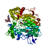
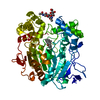
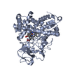
Entry Database : PDB / ID : 6hpuTitle Crystal structure of human Pif1 helicase in complex with ADP-AlF4 ATP-dependent DNA helicase PIF1 Keywords / / / / / Function / homology Function Domain/homology Component
/ / / / / / / / / / / / / / / / / / / / / / / / / / / / / / / / / / / / / / / / / / / / / / / / / / / / Biological species Homo sapiens (human)Method / / / Resolution : 3.96 Å Authors Levdikov, V.M. / Dehghani-Tafti, S. / Bax, B.D. / Sanders, C.M. / Antson, A.A. Journal : Nucleic Acids Res. / Year : 2019Title : Structural and functional analysis of the nucleotide and DNA binding activities of the human PIF1 helicase.Authors : Dehghani-Tafti, S. / Levdikov, V. / Antson, A.A. / Bax, B. / Sanders, C.M. History Deposition Sep 21, 2018 Deposition site / Processing site Revision 1.0 Jan 23, 2019 Provider / Type Revision 1.1 Feb 13, 2019 Group / Database references / Category / citation_author / pdbx_database_procItem _citation.country / _citation.journal_abbrev ... _citation.country / _citation.journal_abbrev / _citation.journal_id_ASTM / _citation.journal_id_CSD / _citation.journal_id_ISSN / _citation.pdbx_database_id_DOI / _citation.pdbx_database_id_PubMed / _citation.year / _citation_author.identifier_ORCID / _citation_author.name Revision 1.2 Apr 17, 2019 Group / Database references / Category / pdbx_database_procItem / _citation.page_first / _citation.page_lastRevision 1.3 Jan 24, 2024 Group / Database references / Refinement descriptionCategory chem_comp_atom / chem_comp_bond ... chem_comp_atom / chem_comp_bond / database_2 / pdbx_initial_refinement_model Item / _database_2.pdbx_database_accession
Show all Show less
 Movie
Movie Controller

 Yorodumi
 Open data
 Basic information
 Components
 Keywords
 Function and homology information
 Homo sapiens (human)
 X-RAY DIFFRACTION /
  Authors
 Citation
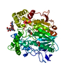
 Structure visualization
 Downloads & links
 Other downloads


 PDBj
PDBj


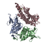
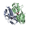
 Assembly


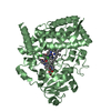
 Mass: 427.201 Da / Num. of mol.: 2 / Source method: obtained synthetically / Formula: C10H15N5O10P2 / Comment: ADP, energy-carrying molecule*YM
 Mass: 102.975 Da / Num. of mol.: 2 / Source method: obtained synthetically / Formula: AlF4
 Mass: 24.305 Da / Num. of mol.: 2 / Source method: obtained synthetically / Formula: Mg
 Sample preparation
 / Beamline: I04 / Wavelength: 0.92819 Å
 Processing