 Movie
Movie Controller


+ Open data

- Basic information
| Entry | Database: PDB / ID: 3wri | ||||||
|---|---|---|---|---|---|---|---|
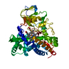
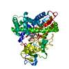
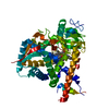
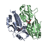
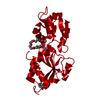
| Title | Crystal structure of P450cam | ||||||
 Components Components | Camphor 5-monooxygenase | ||||||
 Keywords Keywords | OXIDOREDUCTASE / Metal-binding / Heme | ||||||
| Function / homology |  Function and homology information Function and homology informationcamphor 5-monooxygenase / camphor 5-monooxygenase activity / (+)-camphor catabolic process / iron ion binding / heme binding / cytoplasm Similarity search - Function | ||||||
| Biological species |  Pseudomonas putida (bacteria) Pseudomonas putida (bacteria) | ||||||
| Method |  X-RAY DIFFRACTION / SYNCHROTRON / MOLECULAR REPLACEMENT / Resolution: 2.9 Å X-RAY DIFFRACTION / SYNCHROTRON / MOLECULAR REPLACEMENT / Resolution: 2.9 Å | ||||||
 Authors Authors | Kishimoto, A. / Takagi, K. / Amano, A. / Sakurai, K. / Mizushima, T. / Shimada, H. | ||||||
 Citation Citation | Journal: To be Published Title: Structure of P450cam intermediate Authors: Kishimoto, A. / Takagi, K. / Amano, A. / Sakurai, K. / Katayama, Y. / Aminaka, R. / Ito, M. / Mizushima, T. / Shimada, H. | ||||||
| History |
|
- Structure visualization
| Structure viewer | Molecule: MolmilJmol/JSmol |
|---|
- Downloads & links
-Download
| PDBx/mmCIF format | 3wri.cif.gz | 174 KB | Display | PDBx/mmCIF format |
|---|---|---|---|---|
| PDB format | pdb3wri.ent.gz | 137.9 KB | Display | PDB format |
| PDBx/mmJSON format | 3wri.json.gz | Tree view | PDBx/mmJSON format | |
| Others |  Other downloads Other downloads |
-Validation report
| Arichive directory | https://data.pdbj.org/pub/pdb/validation_reports/wr/3wriftp://data.pdbj.org/pub/pdb/validation_reports/wr/3wri | HTTPS FTP |
|---|
-Related structure data
| Related structure data |  3l62S S: Starting model for refinement |
|---|---|
| Similar structure data |


-Links
 PDBj
PDBj
- Assembly
| Deposited unit | 
| ||||||||
|---|---|---|---|---|---|---|---|---|---|
| 1 | 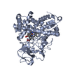
| ||||||||
| 2 | 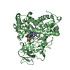
| ||||||||
| Unit cell |
|
-Components
| #1: Protein | Mass: 47548.945 Da / Num. of mol.: 2 Source method: isolated from a genetically manipulated source Source: (gene. exp.) Pseudomonas putida (bacteria) / Gene: camC, cyp101 / Plasmid: PUC18 / Production host: #2: Chemical |   Mass: 616.487 Da / Num. of mol.: 2 / Source method: obtained synthetically / Formula: C34H32FeN4O4 Mass: 616.487 Da / Num. of mol.: 2 / Source method: obtained synthetically / Formula: C34H32FeN4O4#3: Chemical |   Mass: 152.233 Da / Num. of mol.: 2 / Source method: obtained synthetically / Formula: C10H16O Mass: 152.233 Da / Num. of mol.: 2 / Source method: obtained synthetically / Formula: C10H16O#4: Water | ChemComp-HOH / |  Mass: 18.015 Da / Num. of mol.: 9 / Source method: isolated from a natural source / Formula: H2O Mass: 18.015 Da / Num. of mol.: 9 / Source method: isolated from a natural source / Formula: H2O |
|---|
-Experimental details
-Experiment
| Experiment | Method: X-RAY DIFFRACTION / Number of used crystals: 1 |
|---|
- Sample preparation
| Crystal | Density Matthews: 2.22 Å3/Da / Density % sol: 44.62 % |
|---|---|
| Crystal grow | Temperature: 279 K / Method: vapor diffusion, sitting drop / pH: 6.5 Details: 50mM Tris-HCl, 200mM KCl,20-30% PEG 4000, 0.001mM Camphor, pH 6.5, VAPOR DIFFUSION, SITTING DROP, temperature 279K |
-Data collection
| Diffraction | Mean temperature: 100 K |
|---|---|
| Diffraction source | Source: SYNCHROTRON / Site: SPring-8  / Beamline: BL44XU / Wavelength: 0.9 Å / Beamline: BL44XU / Wavelength: 0.9 Å |
| Detector | Type: RAYONIX MX300HE / Detector: CCD / Date: Dec 17, 2012 |
| Radiation | Protocol: SINGLE WAVELENGTH / Monochromatic (M) / Laue (L): M / Scattering type: x-ray |
| Radiation wavelength | Wavelength: 0.9 Å / Relative weight: 1 |
| Reflection | Resolution: 2.9→50 Å / Num. obs: 18663 / % possible obs: 100 % / Observed criterion σ(I): -3 / Redundancy: 3.8 % / Rmerge(I) obs: 0.099 / Net I/σ(I): 14.8 |
| Reflection shell | Resolution: 2.9→2.95 Å / Redundancy: 3.8 % / Rmerge(I) obs: 0.426 / Mean I/σ(I) obs: 5.8 / % possible all: 100 |
- Processing
| Software |
| ||||||||||||||||||||||||||||||||||||||||||||||||||||||||
|---|---|---|---|---|---|---|---|---|---|---|---|---|---|---|---|---|---|---|---|---|---|---|---|---|---|---|---|---|---|---|---|---|---|---|---|---|---|---|---|---|---|---|---|---|---|---|---|---|---|---|---|---|---|---|---|---|---|
| Refinement | Method to determine structure: MOLECULAR REPLACEMENT Starting model: 3L62 Resolution: 2.9→46.318 Å / Cor.coef. Fo:Fc: 0.915 / Cor.coef. Fo:Fc free: 0.819 / SU ML: 0.4 / σ(F): 1.35 / Phase error: 27.35 / Stereochemistry target values: ML / Details: HYDROGENS HAVE BEEN ADDED IN THE RIDING POSITIONS
| ||||||||||||||||||||||||||||||||||||||||||||||||||||||||
| Solvent computation | Shrinkage radii: 1.06 Å / VDW probe radii: 1.3 Å / Solvent model: FLAT BULK SOLVENT MODEL / Bsol: 11.727 Å2 / ksol: 0.278 e/Å3 | ||||||||||||||||||||||||||||||||||||||||||||||||||||||||
| Displacement parameters |
| ||||||||||||||||||||||||||||||||||||||||||||||||||||||||
| Refinement step | Cycle: LAST / Resolution: 2.9→46.318 Å
| ||||||||||||||||||||||||||||||||||||||||||||||||||||||||
| Refine LS restraints |
| ||||||||||||||||||||||||||||||||||||||||||||||||||||||||
| LS refinement shell |
|
