 Movie
Movie Controller

[English] 日本語
 Yorodumi
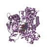
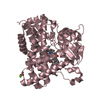
- PDB-3a4z: Structure of cytochrome P450 Vdh mutant (Vdh-K1) obtained by dire... -
+ Open data

- Basic information
| Entry | Database: PDB / ID: 3a4z | ||||||
|---|---|---|---|---|---|---|---|
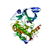
| Title | Structure of cytochrome P450 Vdh mutant (Vdh-K1) obtained by directed evolution | ||||||
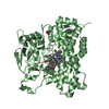
 Components Components | Vitamin D hydroxylase | ||||||
 Keywords Keywords | OXIDOREDUCTASE / Cytochrome P450 / vitamin D3 hydroxylase / hemoprotein / monooxygenase / directed evolution | ||||||
| Function / homology |  Function and homology information Function and homology informationcholestanetriol 26-monooxygenase / cholestanetetraol 26-dehydrogenase activity / iron ion binding / heme binding / cytoplasm Similarity search - Function | ||||||
| Biological species |  Pseudonocardia autotrophica (bacteria) Pseudonocardia autotrophica (bacteria) | ||||||
| Method |  X-RAY DIFFRACTION / SYNCHROTRON / MOLECULAR REPLACEMENT / Resolution: 2.2 Å X-RAY DIFFRACTION / SYNCHROTRON / MOLECULAR REPLACEMENT / Resolution: 2.2 Å | ||||||
 Authors Authors | Yasutake, Y. / Fujii, Y. / Cheon, W.K. / Arisawa, A. / Tamura, T. | ||||||
 Citation Citation | Journal: J.Biol.Chem. / Year: 2010 Title: Structural evidence for enhancement of sequential vitamin D3 hydroxylation activities by directed evolution of cytochrome P450 vitamin D3 hydroxylase Authors: Yasutake, Y. / Fujii, Y. / Nishioka, T. / Cheon, W.K. / Arisawa, A. / Tamura, T. | ||||||
| History |
|
- Structure visualization
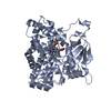
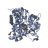
| Structure viewer | Molecule: MolmilJmol/JSmol |
|---|
- Downloads & links
-Download
| PDBx/mmCIF format | 3a4z.cif.gz | 409.5 KB | Display | PDBx/mmCIF format |
|---|---|---|---|---|
| PDB format | pdb3a4z.ent.gz | 334.4 KB | Display | PDB format |
| PDBx/mmJSON format | 3a4z.json.gz | Tree view | PDBx/mmJSON format | |
| Others |  Other downloads Other downloads |
-Validation report
| Arichive directory | https://data.pdbj.org/pub/pdb/validation_reports/a4/3a4zftp://data.pdbj.org/pub/pdb/validation_reports/a4/3a4z | HTTPS FTP |
|---|
-Related structure data
| Related structure data |  3a4gSC  3a4hC  3a50C  3a51C S: Starting model for refinement C: citing same article ( |
|---|---|
| Similar structure data |


-Links
 PDBj
PDBj
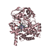
- Assembly
| Deposited unit | 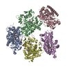
| ||||||||
|---|---|---|---|---|---|---|---|---|---|
| 1 | 
| ||||||||
| 2 | 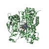
| ||||||||
| 3 | 
| ||||||||
| 4 | 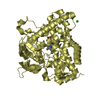
| ||||||||
| 5 | 
| ||||||||
| Unit cell |
|
-Components
-Protein , 1 types, 5 molecules ABCDE
| #1: Protein | Mass: 45587.887 Da / Num. of mol.: 5 / Mutation: T70R, V156L, E216M, E384R Source method: isolated from a genetically manipulated source Source: (gene. exp.) Pseudonocardia autotrophica (bacteria) / Strain: NBRC 12743 / Gene: vdh / Plasmid: pET22 / Production host: |
|---|
-Non-polymers , 5 types, 833 molecules 


| #2: Chemical | ChemComp-HEM /  Mass: 616.487 Da / Num. of mol.: 5 / Source method: obtained synthetically / Formula: C34H32FeN4O4 Mass: 616.487 Da / Num. of mol.: 5 / Source method: obtained synthetically / Formula: C34H32FeN4O4#3: Chemical | ChemComp-CA /  Mass: 40.078 Da / Num. of mol.: 6 / Source method: obtained synthetically / Formula: Ca Mass: 40.078 Da / Num. of mol.: 6 / Source method: obtained synthetically / Formula: Ca#4: Chemical | ChemComp-ACT /  Mass: 59.044 Da / Num. of mol.: 6 / Source method: obtained synthetically / Formula: C2H3O2 Mass: 59.044 Da / Num. of mol.: 6 / Source method: obtained synthetically / Formula: C2H3O2#5: Chemical |  Mass: 92.094 Da / Num. of mol.: 2 / Source method: obtained synthetically / Formula: C3H8O3 Mass: 92.094 Da / Num. of mol.: 2 / Source method: obtained synthetically / Formula: C3H8O3#6: Water | ChemComp-HOH / | Mass: 18.015 Da / Num. of mol.: 814 / Source method: isolated from a natural source / Formula: H2O |
|---|
-Experimental details
-Experiment
| Experiment | Method: X-RAY DIFFRACTION / Number of used crystals: 1 |
|---|
- Sample preparation
| Crystal | Density Matthews: 2.78 Å3/Da / Density % sol: 55.74 % |
|---|---|
| Crystal grow | Temperature: 293 K / Method: vapor diffusion, hanging drop / pH: 7.5 Details: 0.1M calcium acetate, 13% PEG3350, pH7.5, VAPOR DIFFUSION, HANGING DROP, temperature 293K |
-Data collection
| Diffraction | Mean temperature: 100 K |
|---|---|
| Diffraction source | Source: SYNCHROTRON / Site: Photon Factory  / Beamline: AR-NE3A / Wavelength: 1 Å / Beamline: AR-NE3A / Wavelength: 1 Å |
| Detector | Type: ADSC QUANTUM 270 / Detector: CCD / Date: Jun 21, 2009 |
| Radiation | Monochromator: Si(111) double crystal monochromator / Protocol: SINGLE WAVELENGTH / Monochromatic (M) / Laue (L): M / Scattering type: x-ray |
| Radiation wavelength | Wavelength: 1 Å / Relative weight: 1 |
| Reflection | Resolution: 2.2→50 Å / Num. obs: 129009 / % possible obs: 100 % / Redundancy: 7.3 % / Biso Wilson estimate: 27.9 Å2 / Rmerge(I) obs: 0.101 / Net I/σ(I): 26 |
| Reflection shell | Resolution: 2.2→2.24 Å / Redundancy: 6 % / Rmerge(I) obs: 0.499 / Mean I/σ(I) obs: 2.86 / Num. unique all: 6327 / % possible all: 99.8 |
- Processing
| Software |
| ||||||||||||||||||||||||||||||||||||||||||||||||||||||||||||||||||||||||||||||||||||||||||||||||||||
|---|---|---|---|---|---|---|---|---|---|---|---|---|---|---|---|---|---|---|---|---|---|---|---|---|---|---|---|---|---|---|---|---|---|---|---|---|---|---|---|---|---|---|---|---|---|---|---|---|---|---|---|---|---|---|---|---|---|---|---|---|---|---|---|---|---|---|---|---|---|---|---|---|---|---|---|---|---|---|---|---|---|---|---|---|---|---|---|---|---|---|---|---|---|---|---|---|---|---|---|---|---|
| Refinement | Method to determine structure: MOLECULAR REPLACEMENT Starting model: PDB ENTRY 3A4G Resolution: 2.2→49.27 Å / Cor.coef. Fo:Fc: 0.945 / Cor.coef. Fo:Fc free: 0.92 / SU B: 5.627 / SU ML: 0.144 / Cross valid method: THROUGHOUT / ESU R: 0.245 / ESU R Free: 0.202 / Stereochemistry target values: MAXIMUM LIKELIHOOD / Details: HYDROGENS HAVE BEEN ADDED IN THE RIDING POSITIONS
| ||||||||||||||||||||||||||||||||||||||||||||||||||||||||||||||||||||||||||||||||||||||||||||||||||||
| Solvent computation | Ion probe radii: 0.8 Å / Shrinkage radii: 0.8 Å / VDW probe radii: 1.2 Å / Solvent model: MASK | ||||||||||||||||||||||||||||||||||||||||||||||||||||||||||||||||||||||||||||||||||||||||||||||||||||
| Displacement parameters | Biso mean: 34.106 Å2
| ||||||||||||||||||||||||||||||||||||||||||||||||||||||||||||||||||||||||||||||||||||||||||||||||||||
| Refinement step | Cycle: LAST / Resolution: 2.2→49.27 Å
| ||||||||||||||||||||||||||||||||||||||||||||||||||||||||||||||||||||||||||||||||||||||||||||||||||||
| Refine LS restraints |
| ||||||||||||||||||||||||||||||||||||||||||||||||||||||||||||||||||||||||||||||||||||||||||||||||||||
| LS refinement shell | Resolution: 2.202→2.259 Å / Total num. of bins used: 20
|
