 Movie
Movie Controller


+ Open data

- Basic information
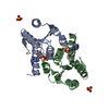
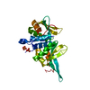
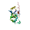
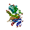
| Entry | Database: PDB / ID: 4kxl | ||||||
|---|---|---|---|---|---|---|---|
| Title | Crystal structure of DNPH1 (RCL) with 6-CYCLOPENTYL-AMP | ||||||
 Components Components | 2'-deoxynucleoside 5'-phosphate N-hydrolase 1 | ||||||
 Keywords Keywords | HYDROLASE / DEOXYRIBONUCLEOSIDE 5'-MONOPHOSPHATE N-GLYCOSIDASE | ||||||
| Function / homology |  Function and homology information Function and homology informationPurine catabolism / 5-hydroxymethyl-dUMP N-hydrolase activity / nucleoside salvage / deoxyribonucleoside monophosphate catabolic process / dGMP catabolic process / allantoin metabolic process / Hydrolases; Glycosylases; Hydrolysing N-glycosyl compounds / epithelial cell differentiation / positive regulation of cell growth / protein homodimerization activity ...Purine catabolism / 5-hydroxymethyl-dUMP N-hydrolase activity / nucleoside salvage / deoxyribonucleoside monophosphate catabolic process / dGMP catabolic process / allantoin metabolic process / Hydrolases; Glycosylases; Hydrolysing N-glycosyl compounds / epithelial cell differentiation / positive regulation of cell growth / protein homodimerization activity / identical protein binding / nucleus / cytoplasm Similarity search - Function | ||||||
| Biological species |  | ||||||
| Method |  X-RAY DIFFRACTION / SYNCHROTRON / MOLECULAR REPLACEMENT / molecular replacement / Resolution: 1.69 Å X-RAY DIFFRACTION / SYNCHROTRON / MOLECULAR REPLACEMENT / molecular replacement / Resolution: 1.69 Å | ||||||
 Authors Authors | Padilla, A. / Labesse, G. / Kaminski, P.A. | ||||||
 Citation Citation | Journal: Plos One / Year: 2013 Title: N (6)-substituted AMPs inhibit mammalian deoxynucleotide N-hydrolase DNPH1. Authors: Amiable, C. / Pochet, S. / Padilla, A. / Labesse, G. / Kaminski, P.A. #1: Journal: Acta Crystallogr.,Sect.D / Year: 2013Title: Structure of the oncoprotein Rcl bound to three nucleotide analogues. Authors: Padilla, A. / Amiable, C. / Pochet, S. / Kaminski, P.A. / Labesse, G. | ||||||
| History |
|
- Structure visualization
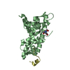
| Structure viewer | Molecule: MolmilJmol/JSmol |
|---|
- Downloads & links
-Download
| PDBx/mmCIF format | 4kxl.cif.gz | 250.3 KB | Display | PDBx/mmCIF format |
|---|---|---|---|---|
| PDB format | pdb4kxl.ent.gz | 201.3 KB | Display | PDB format |
| PDBx/mmJSON format | 4kxl.json.gz | Tree view | PDBx/mmJSON format | |
| Others |  Other downloads Other downloads |
-Validation report
| Arichive directory | https://data.pdbj.org/pub/pdb/validation_reports/kx/4kxlftp://data.pdbj.org/pub/pdb/validation_reports/kx/4kxl | HTTPS FTP |
|---|
-Related structure data
| Related structure data | 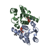 4kxmC  4kxnC  4fyiS S: Starting model for refinement C: citing same article ( |
|---|---|
| Similar structure data |

-Links
 PDBj
PDBj- Assembly
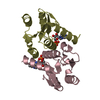
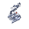
| Deposited unit | 
| ||||||||
|---|---|---|---|---|---|---|---|---|---|
| 1 | 
| ||||||||
| 2 | 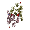
| ||||||||
| Unit cell |
|
-Components
| #1: Protein | Mass: 17173.334 Da / Num. of mol.: 4 / Fragment: UNP residues 11-151 / Mutation: D69N Source method: isolated from a genetically manipulated source Source: (gene. exp.)  References: UniProt: O35820, Hydrolases; Glycosylases; Hydrolysing N-glycosyl compounds #2: Chemical | ChemComp-6C6 /   Mass: 415.338 Da / Num. of mol.: 4 / Source method: obtained synthetically / Formula: C15H22N5O7P Mass: 415.338 Da / Num. of mol.: 4 / Source method: obtained synthetically / Formula: C15H22N5O7P#3: Chemical |   Mass: 96.063 Da / Num. of mol.: 2 / Source method: obtained synthetically / Formula: SO4 Mass: 96.063 Da / Num. of mol.: 2 / Source method: obtained synthetically / Formula: SO4#4: Water | ChemComp-HOH / |  Mass: 18.015 Da / Num. of mol.: 497 / Source method: isolated from a natural source / Formula: H2O Mass: 18.015 Da / Num. of mol.: 497 / Source method: isolated from a natural source / Formula: H2O |
|---|
-Experimental details
-Experiment
| Experiment | Method: X-RAY DIFFRACTION / Number of used crystals: 1 |
|---|
- Sample preparation
| Crystal | Density Matthews: 1.87 Å3/Da / Density % sol: 34.23 % |
|---|---|
| Crystal grow | Temperature: 291 K / Method: hanging drop / pH: 7.6 Details: 100 MM TRIS, 1.1 AMMONIUM SULPHATE, 20MM MGSO4, PH 7.6, hanging drop, temperature 291K |
-Data collection
| Diffraction source | Source: SYNCHROTRON / Site: ESRF  / Beamline: ID14-4 / Wavelength: 0.9795 Å / Beamline: ID14-4 / Wavelength: 0.9795 Å |
|---|---|
| Detector | Type: ADSC QUANTUM 315r / Detector: CCD / Date: Jul 25, 2010 |
| Radiation | Monochromator: CHANNEL CUT ESRF MONOCHROMATOR / Protocol: SINGLE WAVELENGTH / Monochromatic (M) / Laue (L): M / Scattering type: x-ray |
| Radiation wavelength | Wavelength: 0.9795 Å / Relative weight: 1 |
| Reflection | Resolution: 1.69→32.03 Å / Num. obs: 53250 / % possible obs: 93.88 % / Observed criterion σ(I): 1 / Redundancy: 3.9 % / Rmerge(I) obs: 0.065 / Net I/σ(I): 5.4 |
| Reflection shell | Resolution: 1.69→1.79 Å / Redundancy: 2.8 % / Rmerge(I) obs: 0.115 / Mean I/σ(I) obs: 2.6 / % possible all: 75.8 |
-Phasing
| Phasing | Method: molecular replacement |
|---|
- Processing
| Software |
| ||||||||||||||||||||||||||||||||||||||||||||||||||||||||||||||||||||||||||||||||||||||||||||||||||||||||||||||||||||||||||||||||||||||||||||||||||||||
|---|---|---|---|---|---|---|---|---|---|---|---|---|---|---|---|---|---|---|---|---|---|---|---|---|---|---|---|---|---|---|---|---|---|---|---|---|---|---|---|---|---|---|---|---|---|---|---|---|---|---|---|---|---|---|---|---|---|---|---|---|---|---|---|---|---|---|---|---|---|---|---|---|---|---|---|---|---|---|---|---|---|---|---|---|---|---|---|---|---|---|---|---|---|---|---|---|---|---|---|---|---|---|---|---|---|---|---|---|---|---|---|---|---|---|---|---|---|---|---|---|---|---|---|---|---|---|---|---|---|---|---|---|---|---|---|---|---|---|---|---|---|---|---|---|---|---|---|---|---|---|---|
| Refinement | Method to determine structure: MOLECULAR REPLACEMENT Starting model: PDB ENTRY 4FYI Resolution: 1.69→27.02 Å / Cor.coef. Fo:Fc: 0.949 / Cor.coef. Fo:Fc free: 0.927 / Occupancy max: 1 / Occupancy min: 0.2 / SU B: 4.665 / SU ML: 0.072 / Cross valid method: THROUGHOUT / σ(F): 0 / ESU R: 0.123 / ESU R Free: 0.116 / Stereochemistry target values: MAXIMUM LIKELIHOOD Details: HYDROGENS HAVE BEEN ADDED IN THE RIDING POSITIONS U VALUES : WITH TLS ADDED
| ||||||||||||||||||||||||||||||||||||||||||||||||||||||||||||||||||||||||||||||||||||||||||||||||||||||||||||||||||||||||||||||||||||||||||||||||||||||
| Solvent computation | Ion probe radii: 0.8 Å / Shrinkage radii: 0.8 Å / VDW probe radii: 1.4 Å / Solvent model: MASK | ||||||||||||||||||||||||||||||||||||||||||||||||||||||||||||||||||||||||||||||||||||||||||||||||||||||||||||||||||||||||||||||||||||||||||||||||||||||
| Displacement parameters | Biso max: 59.06 Å2 / Biso mean: 16.691 Å2 / Biso min: 4.48 Å2
| ||||||||||||||||||||||||||||||||||||||||||||||||||||||||||||||||||||||||||||||||||||||||||||||||||||||||||||||||||||||||||||||||||||||||||||||||||||||
| Refinement step | Cycle: LAST / Resolution: 1.69→27.02 Å
| ||||||||||||||||||||||||||||||||||||||||||||||||||||||||||||||||||||||||||||||||||||||||||||||||||||||||||||||||||||||||||||||||||||||||||||||||||||||
| Refine LS restraints |
| ||||||||||||||||||||||||||||||||||||||||||||||||||||||||||||||||||||||||||||||||||||||||||||||||||||||||||||||||||||||||||||||||||||||||||||||||||||||
| LS refinement shell | Resolution: 1.69→1.734 Å / Total num. of bins used: 20
| ||||||||||||||||||||||||||||||||||||||||||||||||||||||||||||||||||||||||||||||||||||||||||||||||||||||||||||||||||||||||||||||||||||||||||||||||||||||
| Refinement TLS params. | Method: refined / Refine-ID: X-RAY DIFFRACTION
| ||||||||||||||||||||||||||||||||||||||||||||||||||||||||||||||||||||||||||||||||||||||||||||||||||||||||||||||||||||||||||||||||||||||||||||||||||||||
| Refinement TLS group |
|
