 Movie
Movie Controller

[English] 日本語
 Yorodumi
- PDB-4kfc: Crystal structure of a hyperactive mutant of response regulator K... -
+ Open data

- Basic information
| Entry | Database: PDB / ID: 4kfc | ||||||
|---|---|---|---|---|---|---|---|
| Title | Crystal structure of a hyperactive mutant of response regulator KdpE complexed to its promoter DNA | ||||||
 Components Components |
| ||||||
 Keywords Keywords | TRANSCRIPTION REGULATOR/DNA / receiver domain / DNA-binding domain / TRANSCRIPTION REGULATOR-DNA complex | ||||||
| Function / homology |  Function and homology information Function and homology informationphosphorelay response regulator activity / DNA-binding transcription activator activity / cis-regulatory region sequence-specific DNA binding / protein-DNA complex / transcription cis-regulatory region binding / DNA-binding transcription factor activity / regulation of DNA-templated transcription / protein homodimerization activity / cytosol Similarity search - Function | ||||||
| Biological species |  | ||||||
| Method |  X-RAY DIFFRACTION / SYNCHROTRON / MOLECULAR REPLACEMENT / Resolution: 2.53 Å X-RAY DIFFRACTION / SYNCHROTRON / MOLECULAR REPLACEMENT / Resolution: 2.53 Å | ||||||
 Authors Authors | Kumar, S. / Narayanan, A. / Yernool, D.A. | ||||||
 Citation Citation | Journal: Nat Commun / Year: 2014 Title: An asymmetric heterodomain interface stabilizes a response regulator-DNA complex. Authors: Narayanan, A. / Kumar, S. / Evrard, A.N. / Paul, L.N. / Yernool, D.A. | ||||||
| History |
|
- Structure visualization
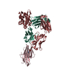
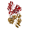
| Structure viewer | Molecule: MolmilJmol/JSmol |
|---|
- Downloads & links
-Download
| PDBx/mmCIF format | 4kfc.cif.gz | 138.5 KB | Display | PDBx/mmCIF format |
|---|---|---|---|---|
| PDB format | pdb4kfc.ent.gz | 103.9 KB | Display | PDB format |
| PDBx/mmJSON format | 4kfc.json.gz | Tree view | PDBx/mmJSON format | |
| Others |  Other downloads Other downloads |
-Validation report
| Arichive directory | https://data.pdbj.org/pub/pdb/validation_reports/kf/4kfcftp://data.pdbj.org/pub/pdb/validation_reports/kf/4kfc | HTTPS FTP |
|---|
-Related structure data
| Related structure data | 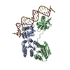 4knyC 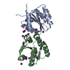 4l85C 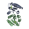 1zh4S 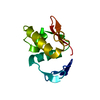 3zq7S S: Starting model for refinement C: citing same article ( |
|---|---|
| Similar structure data |

-Links
 PDBj
PDBj


- Assembly
| Deposited unit | 
| ||||||||
|---|---|---|---|---|---|---|---|---|---|
| 1 |
| ||||||||
| Unit cell |
| ||||||||
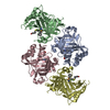
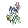
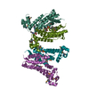
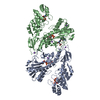
| Details | The present structures with two protein molecules bound to dsDNA, represents the biological assemble. |
-Components
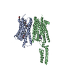
| #1: Protein | Mass: 25437.207 Da / Num. of mol.: 2 / Fragment: UNP residues 3-225 / Mutation: E216A Source method: isolated from a genetically manipulated source Source: (gene. exp.) #2: DNA chain | | Mass: 9025.812 Da / Num. of mol.: 1 / Source method: obtained synthetically / Details: CHEMICALLY SYNTHESIZED #3: DNA chain | | Mass: 9418.117 Da / Num. of mol.: 1 / Source method: obtained synthetically / Details: CHEMICALLY SYNTHESIZED #4: Water | ChemComp-HOH / |  Mass: 18.015 Da / Num. of mol.: 148 / Source method: isolated from a natural source / Formula: H2O Mass: 18.015 Da / Num. of mol.: 148 / Source method: isolated from a natural source / Formula: H2O |
|---|
-Experimental details
-Experiment
| Experiment | Method: X-RAY DIFFRACTION / Number of used crystals: 1 |
|---|
- Sample preparation
| Crystal | Density Matthews: 4.28 Å3/Da / Density % sol: 71.28 % |
|---|---|
| Crystal grow | Temperature: 293 K / Method: vapor diffusion, hanging drop / pH: 5.5 Details: 3-5% PEG 4K, 0.1 M sodium acetate buffer (pH 5.5), 0.1 M MgCl2, 2mM L-proline, VAPOR DIFFUSION, HANGING DROP, temperature 293K |
-Data collection
| Diffraction | Mean temperature: 100 K |
|---|---|
| Diffraction source | Source: SYNCHROTRON / Site: APS  / Beamline: 23-ID-B / Wavelength: 1.0332 Å / Beamline: 23-ID-B / Wavelength: 1.0332 Å |
| Detector | Type: MARMOSAIC 300 mm CCD / Detector: CCD / Date: Oct 30, 2011 |
| Radiation | Protocol: SINGLE WAVELENGTH / Monochromatic (M) / Laue (L): M / Scattering type: x-ray |
| Radiation wavelength | Wavelength: 1.0332 Å / Relative weight: 1 |
| Reflection | Resolution: 2.53→44.43 Å / Num. all: 40355 / Num. obs: 40336 / % possible obs: 99.95 % / Observed criterion σ(F): 0 / Observed criterion σ(I): 0 / Redundancy: 12 % / Rmerge(I) obs: 0.11 / Net I/σ(I): 13.48 |
| Reflection shell | Resolution: 2.53→2.62 Å / Redundancy: 12.2 % / Rmerge(I) obs: 0.49 / Mean I/σ(I) obs: 4.4 / Num. unique all: 40355 / % possible all: 100 |
- Processing
| Software |
| ||||||||||||||||||||||||||||||||||||||||||||||||||||||||||||||||||||||||||||||||||||||||||||||||||||||||||||||||||||||||||||||||||||||||||||||||||||||||||||||||||||||||||||||||||||||||||||||||||||||||||||||||||
|---|---|---|---|---|---|---|---|---|---|---|---|---|---|---|---|---|---|---|---|---|---|---|---|---|---|---|---|---|---|---|---|---|---|---|---|---|---|---|---|---|---|---|---|---|---|---|---|---|---|---|---|---|---|---|---|---|---|---|---|---|---|---|---|---|---|---|---|---|---|---|---|---|---|---|---|---|---|---|---|---|---|---|---|---|---|---|---|---|---|---|---|---|---|---|---|---|---|---|---|---|---|---|---|---|---|---|---|---|---|---|---|---|---|---|---|---|---|---|---|---|---|---|---|---|---|---|---|---|---|---|---|---|---|---|---|---|---|---|---|---|---|---|---|---|---|---|---|---|---|---|---|---|---|---|---|---|---|---|---|---|---|---|---|---|---|---|---|---|---|---|---|---|---|---|---|---|---|---|---|---|---|---|---|---|---|---|---|---|---|---|---|---|---|---|---|---|---|---|---|---|---|---|---|---|---|---|---|---|---|---|---|
| Refinement | Method to determine structure: MOLECULAR REPLACEMENT Starting model: 1ZH4 and 3ZQ7 Resolution: 2.53→44.32 Å / SU ML: 0.31 / σ(F): 1.33 / Phase error: 25.02 / Stereochemistry target values: ML
| ||||||||||||||||||||||||||||||||||||||||||||||||||||||||||||||||||||||||||||||||||||||||||||||||||||||||||||||||||||||||||||||||||||||||||||||||||||||||||||||||||||||||||||||||||||||||||||||||||||||||||||||||||
| Solvent computation | Shrinkage radii: 0.9 Å / VDW probe radii: 1.11 Å / Solvent model: FLAT BULK SOLVENT MODEL | ||||||||||||||||||||||||||||||||||||||||||||||||||||||||||||||||||||||||||||||||||||||||||||||||||||||||||||||||||||||||||||||||||||||||||||||||||||||||||||||||||||||||||||||||||||||||||||||||||||||||||||||||||
| Refinement step | Cycle: LAST / Resolution: 2.53→44.32 Å
| ||||||||||||||||||||||||||||||||||||||||||||||||||||||||||||||||||||||||||||||||||||||||||||||||||||||||||||||||||||||||||||||||||||||||||||||||||||||||||||||||||||||||||||||||||||||||||||||||||||||||||||||||||
| Refine LS restraints |
| ||||||||||||||||||||||||||||||||||||||||||||||||||||||||||||||||||||||||||||||||||||||||||||||||||||||||||||||||||||||||||||||||||||||||||||||||||||||||||||||||||||||||||||||||||||||||||||||||||||||||||||||||||
| LS refinement shell |
|
