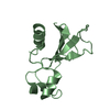
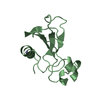
Entry Database : PDB / ID : 3zq7Title The Structure of DNA-binding domain of response regulator from Escherichia coli K-12 KDP OPERON TRANSCRIPTIONAL REGULATORY PROTEIN KDPE Keywords / Function / homology Function Domain/homology Component
/ / / / / / / / / / / / / / / / / / / / / / / / / / / / / / / Biological species ESCHERICHIA COLI (E. coli)Method / / Resolution : 2.52 Å Authors Patil, D.N. / Tomar, S. / Kumar, P. Journal : Plos One / Year : 2012Title : Structure-Function Studies of DNA Binding Domain of Response Regulator Kdpe Reveals Equal Affinity Interactions at DNA Half-Sites.Authors : Narayanan, A. / Paul, L.N. / Tomar, S. / Patil, D.N. / Kumar, P. / Yernool, D.A. History Deposition Jun 8, 2011 Deposition site / Processing site Revision 1.0 Mar 21, 2012 Provider / Type Revision 1.1 Jul 5, 2017 Group / Category / Item Revision 1.2 Dec 20, 2023 Group Data collection / Database references ... Data collection / Database references / Other / Refinement description Category chem_comp_atom / chem_comp_bond ... chem_comp_atom / chem_comp_bond / database_2 / pdbx_database_status / pdbx_initial_refinement_model Item / _database_2.pdbx_database_accession / _pdbx_database_status.status_code_sf
Show all Show less
 Movie
Movie Controller

 Yorodumi
 Open data
 Basic information
 Components
 Keywords
 Function and homology information
 X-RAY DIFFRACTION /
  Authors
 Citation
 Structure visualization
 Downloads & links
 Other downloads


 PDBj
PDBj

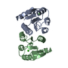
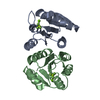
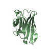
 Assembly
 Mass: 18.015 Da / Num. of mol.: 23 / Source method: isolated from a natural source / Formula: H2O
 Sample preparation
 Processing