 Movie
Movie Controller


+ Open data

- Basic information
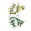
| Entry | Database: PDB / ID: 4k8m | ||||||
|---|---|---|---|---|---|---|---|
| Title | High resolution structure of M.tb NRDH | ||||||
 Components Components | Glutaredoxin-like protein NrdH | ||||||
 Keywords Keywords | ELECTRON TRANSPORT / THIOREDOXIN / REDOXIN / THIOREDOXIN REDUCTASE / RIBONUCLEOTIDE REDUCTASE | ||||||
| Function / homology |  Function and homology information Function and homology information | ||||||
| Biological species |   Mycobacterium tuberculosis (bacteria) Mycobacterium tuberculosis (bacteria) | ||||||
| Method |  X-RAY DIFFRACTION / SYNCHROTRON / MOLECULAR REPLACEMENT / Resolution: 0.87 Å X-RAY DIFFRACTION / SYNCHROTRON / MOLECULAR REPLACEMENT / Resolution: 0.87 Å | ||||||
 Authors Authors | Phulera, S. / Mande, S.C. | ||||||
 Citation Citation | Journal: Biochemistry / Year: 2013 Title: The crystal structure of Mycobacterium tuberculosis NrdH at 0.87 angstrom suggests a possible mode of its activity. Authors: Phulera, S. / Mande, S.C. | ||||||
| History |
|
- Structure visualization
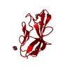
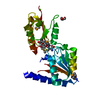
| Structure viewer | Molecule: MolmilJmol/JSmol |
|---|
- Downloads & links
-Download
| PDBx/mmCIF format | 4k8m.cif.gz | 57.1 KB | Display | PDBx/mmCIF format |
|---|---|---|---|---|
| PDB format | pdb4k8m.ent.gz | 40.9 KB | Display | PDB format |
| PDBx/mmJSON format | 4k8m.json.gz | Tree view | PDBx/mmJSON format | |
| Others |  Other downloads Other downloads |
-Validation report
| Arichive directory | https://data.pdbj.org/pub/pdb/validation_reports/k8/4k8mftp://data.pdbj.org/pub/pdb/validation_reports/k8/4k8m | HTTPS FTP |
|---|
-Related structure data
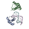
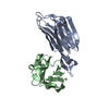
| Related structure data | 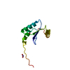 4f2iSC 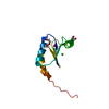 4hs1C S: Starting model for refinement C: citing same article ( |
|---|---|
| Similar structure data |


-Links
 PDBj
PDBj
- Assembly
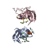
| Deposited unit | 
| ||||||||
|---|---|---|---|---|---|---|---|---|---|
| 1 | 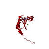
| ||||||||
| Unit cell |
|
-Components
-Protein , 1 types, 1 molecules A
| #1: Protein | Mass: 9125.329 Da / Num. of mol.: 1 Source method: isolated from a genetically manipulated source Source: (gene. exp.) Mycobacterium tuberculosis (bacteria) / Strain: H37RV / Gene: Rv3053c, RVBD_3053c / Plasmid: PET / Production host: |
|---|
-Non-polymers , 5 types, 104 molecules 


| #2: Chemical |  Mass: 62.005 Da / Num. of mol.: 2 / Source method: obtained synthetically / Formula: NO3 Mass: 62.005 Da / Num. of mol.: 2 / Source method: obtained synthetically / Formula: NO3#3: Chemical | ChemComp-CL / |  Mass: 35.453 Da / Num. of mol.: 1 / Source method: obtained synthetically / Formula: Cl Mass: 35.453 Da / Num. of mol.: 1 / Source method: obtained synthetically / Formula: Cl#4: Chemical | ChemComp-GOL / |  Mass: 92.094 Da / Num. of mol.: 1 / Source method: obtained synthetically / Formula: C3H8O3 Mass: 92.094 Da / Num. of mol.: 1 / Source method: obtained synthetically / Formula: C3H8O3#5: Chemical | ChemComp-PEG / |  Mass: 106.120 Da / Num. of mol.: 1 / Source method: obtained synthetically / Formula: C4H10O3 Mass: 106.120 Da / Num. of mol.: 1 / Source method: obtained synthetically / Formula: C4H10O3#6: Water | ChemComp-HOH / | Mass: 18.015 Da / Num. of mol.: 99 / Source method: isolated from a natural source / Formula: H2O |
|---|
-Details
| Has protein modification | Y |
|---|
-Experimental details
-Experiment
| Experiment | Method: X-RAY DIFFRACTION / Number of used crystals: 1 |
|---|
- Sample preparation
| Crystal | Density Matthews: 1.76 Å3/Da / Density % sol: 29.98 % |
|---|
-Data collection
| Diffraction | Mean temperature: 100 K |
|---|---|
| Diffraction source | Source: SYNCHROTRON / Site: ESRF  / Beamline: BM14 / Wavelength: 0.71255 Å / Beamline: BM14 / Wavelength: 0.71255 Å |
| Detector | Type: MARCCD 225X225 / Detector: CCD / Date: Jul 27, 2012 / Details: MIRRORS |
| Radiation | Monochromator: Band pass 1.9x10⁻⁴ for a Si(111) monochromator Protocol: SINGLE WAVELENGTH / Monochromatic (M) / Laue (L): M / Scattering type: x-ray |
| Radiation wavelength | Wavelength: 0.71255 Å / Relative weight: 1 |
| Reflection | Resolution: 0.87→23.85 Å / Num. all: 53538 / Num. obs: 53538 / % possible obs: 99.8 % / Observed criterion σ(F): 1.9 / Observed criterion σ(I): 1.9 / Redundancy: 9.2 % / Biso Wilson estimate: 5.9 Å2 / Rmerge(I) obs: 0.064 / Net I/σ(I): 16.4 |
- Processing
| Software |
| ||||||||||||||||
|---|---|---|---|---|---|---|---|---|---|---|---|---|---|---|---|---|---|
| Refinement | Method to determine structure: MOLECULAR REPLACEMENT Starting model: 4F2I Resolution: 0.87→23.85 Å / σ(F): 1.35
| ||||||||||||||||
| Refinement step | Cycle: LAST / Resolution: 0.87→23.85 Å
|
