 Movie
Movie Controller

[English] 日本語
 Yorodumi
- PDB-4k65: Structure of an airborne transmissible avian influenza H5 hemaggl... -
+ Open data

- Basic information
| Entry | Database: PDB / ID: 4k65 | ||||||
|---|---|---|---|---|---|---|---|
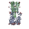
| Title | Structure of an airborne transmissible avian influenza H5 hemagglutinin mutant from the influenza virus A/Indonesia/5/2005 | ||||||
 Components Components | (Hemagglutinin) x 2 | ||||||
 Keywords Keywords | VIRAL PROTEIN / virus attachment / membrane fusion | ||||||
| Function / homology |  Function and homology information Function and homology informationclathrin-dependent endocytosis of virus by host cell / host cell surface receptor binding / fusion of virus membrane with host plasma membrane / fusion of virus membrane with host endosome membrane / viral envelope / virion attachment to host cell / host cell plasma membrane / virion membrane Similarity search - Function | ||||||
| Biological species |   Influenza A virus Influenza A virus | ||||||
| Method |  X-RAY DIFFRACTION / SYNCHROTRON / MOLECULAR REPLACEMENT / Resolution: 2.9 Å X-RAY DIFFRACTION / SYNCHROTRON / MOLECULAR REPLACEMENT / Resolution: 2.9 Å | ||||||
 Authors Authors | Zhang, W. / Shi, Y. / Lu, X. / Shu, Y. / Qi, J. / Gao, G.F. | ||||||
 Citation Citation | Journal: Science / Year: 2013 Title: An airborne transmissible avian influenza H5 hemagglutinin seen at the atomic level. Authors: Zhang, W. / Shi, Y. / Lu, X. / Shu, Y. / Qi, J. / Gao, G.F. | ||||||
| History |
|
- Structure visualization
| Structure viewer | Molecule: MolmilJmol/JSmol |
|---|
- Downloads & links
-Download
| PDBx/mmCIF format | 4k65.cif.gz | 701.7 KB | Display | PDBx/mmCIF format |
|---|---|---|---|---|
| PDB format | pdb4k65.ent.gz | 580.1 KB | Display | PDB format |
| PDBx/mmJSON format | 4k65.json.gz | Tree view | PDBx/mmJSON format | |
| Others |  Other downloads Other downloads |
-Validation report
| Arichive directory | https://data.pdbj.org/pub/pdb/validation_reports/k6/4k65ftp://data.pdbj.org/pub/pdb/validation_reports/k6/4k65 | HTTPS FTP |
|---|
-Related structure data
| Related structure data |  4k62C  4k63C  4k64C  4k66C  4k67C  2fk0S C: citing same article ( S: Starting model for refinement |
|---|---|
| Similar structure data |


-Links
 PDBj
PDBj


- Assembly
| Deposited unit | 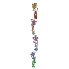
| ||||||||
|---|---|---|---|---|---|---|---|---|---|
| 1 | 
| ||||||||
| 2 | 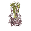
| ||||||||
| 3 | 
| ||||||||
| 4 | 
| ||||||||
| Unit cell |
|
-Components
| #1: Protein | Mass: 36203.953 Da / Num. of mol.: 4 / Fragment: UNP residues 17-326 / Mutation: H107Y, T160A, Q226L, G228S Source method: isolated from a genetically manipulated source Source: (gene. exp.) Influenza A virus / Strain: A/Indonesia/5/2005(H5N1) / Gene: HA / Plasmid: pFastBac1 / Cell line (production host): Hi5 / Production host:  TRICHOPLUSIA NI (cabbage looper) / References: UniProt: A8HWY8 TRICHOPLUSIA NI (cabbage looper) / References: UniProt: A8HWY8#2: Protein | Mass: 18910.838 Da / Num. of mol.: 4 / Fragment: UNP residues 347-510 Source method: isolated from a genetically manipulated source Source: (gene. exp.) Influenza A virus / Strain: A/Indonesia/5/2005(H5N1) / Gene: HA / Plasmid: pFastBac1 / Cell line (production host): Hi5 / Production host: TRICHOPLUSIA NI (cabbage looper) / References: UniProt: A8HWY8#3: Sugar |   Type: D-saccharide, beta linking / Mass: 221.208 Da / Num. of mol.: 2 Type: D-saccharide, beta linking / Mass: 221.208 Da / Num. of mol.: 2Source method: isolated from a genetically manipulated source Formula: C8H15NO6 Has protein modification | Y | |
|---|
-Experimental details
-Experiment
| Experiment | Method: X-RAY DIFFRACTION / Number of used crystals: 1 |
|---|
- Sample preparation
| Crystal | Density Matthews: 3.19 Å3/Da / Density % sol: 61.39 % |
|---|---|
| Crystal grow | Temperature: 291 K / Method: vapor diffusion, sitting drop / pH: 8.5 Details: 25% PEG 1500, 0.1M Tris, pH 8.5, VAPOR DIFFUSION, SITTING DROP, temperature 291K |
-Data collection
| Diffraction | Mean temperature: 100 K |
|---|---|
| Diffraction source | Source: SYNCHROTRON / Site: SSRF  / Beamline: BL17U / Wavelength: 1 Å / Beamline: BL17U / Wavelength: 1 Å |
| Detector | Type: ADSC QUANTUM 315r / Detector: CCD / Date: Oct 3, 2012 |
| Radiation | Monochromator: GRAPHITE / Protocol: SINGLE WAVELENGTH / Monochromatic (M) / Laue (L): M / Scattering type: x-ray |
| Radiation wavelength | Wavelength: 1 Å / Relative weight: 1 |
| Reflection twin | Type: merohedral / Operator: -h,-k,l / Fraction: 0.431 |
| Reflection | Resolution: 2.9→50 Å / Num. all: 56188 / Num. obs: 56188 / % possible obs: 92.6 % / Observed criterion σ(F): 0 / Observed criterion σ(I): -3 |
| Reflection shell | Resolution: 2.9→3 Å / % possible all: 91.4 |
- Processing
| Software |
| ||||||||||||||||||||||||||||||||||||||||||||||||||||||||||||||||||||||||||||||||||||||||||||||||||||||||||||||||||||||||||||||
|---|---|---|---|---|---|---|---|---|---|---|---|---|---|---|---|---|---|---|---|---|---|---|---|---|---|---|---|---|---|---|---|---|---|---|---|---|---|---|---|---|---|---|---|---|---|---|---|---|---|---|---|---|---|---|---|---|---|---|---|---|---|---|---|---|---|---|---|---|---|---|---|---|---|---|---|---|---|---|---|---|---|---|---|---|---|---|---|---|---|---|---|---|---|---|---|---|---|---|---|---|---|---|---|---|---|---|---|---|---|---|---|---|---|---|---|---|---|---|---|---|---|---|---|---|---|---|---|
| Refinement | Method to determine structure: MOLECULAR REPLACEMENT Starting model: 2FK0 Resolution: 2.9→48.876 Å / Occupancy max: 1 / Occupancy min: 1 / FOM work R set: 0.8088 / SU ML: 0.34 / σ(F): 1.97 / Phase error: 26.84 / Stereochemistry target values: ML
| ||||||||||||||||||||||||||||||||||||||||||||||||||||||||||||||||||||||||||||||||||||||||||||||||||||||||||||||||||||||||||||||
| Solvent computation | Shrinkage radii: 0.9 Å / VDW probe radii: 1.11 Å / Solvent model: FLAT BULK SOLVENT MODEL | ||||||||||||||||||||||||||||||||||||||||||||||||||||||||||||||||||||||||||||||||||||||||||||||||||||||||||||||||||||||||||||||
| Displacement parameters | Biso max: 185.66 Å2 / Biso mean: 61.2666 Å2 / Biso min: 2.34 Å2 | ||||||||||||||||||||||||||||||||||||||||||||||||||||||||||||||||||||||||||||||||||||||||||||||||||||||||||||||||||||||||||||||
| Refinement step | Cycle: LAST / Resolution: 2.9→48.876 Å
| ||||||||||||||||||||||||||||||||||||||||||||||||||||||||||||||||||||||||||||||||||||||||||||||||||||||||||||||||||||||||||||||
| Refine LS restraints |
| ||||||||||||||||||||||||||||||||||||||||||||||||||||||||||||||||||||||||||||||||||||||||||||||||||||||||||||||||||||||||||||||
| LS refinement shell | Refine-ID: X-RAY DIFFRACTION / Total num. of bins used: 20
| ||||||||||||||||||||||||||||||||||||||||||||||||||||||||||||||||||||||||||||||||||||||||||||||||||||||||||||||||||||||||||||||
| Refinement TLS params. | Method: refined / Origin x: 24.5671 Å / Origin y: -2.3255 Å / Origin z: -174.9651 Å
| ||||||||||||||||||||||||||||||||||||||||||||||||||||||||||||||||||||||||||||||||||||||||||||||||||||||||||||||||||||||||||||||
| Refinement TLS group |
|
