 Movie
Movie Controller

[English] 日本語
 Yorodumi
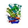
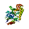
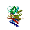
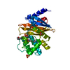
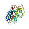
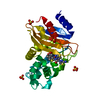
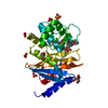
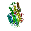
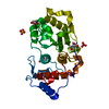
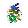
- PDB-4jpm: Structure of SHV-1 beta-lactamase in complex with the 7-alkyliden... -
+ Open data

- Basic information
| Entry | Database: PDB / ID: 4jpm | ||||||
|---|---|---|---|---|---|---|---|
| Title | Structure of SHV-1 beta-lactamase in complex with the 7-alkylidenecephalosporin DCM-1-10 at 1.14 Ang resolution | ||||||
 Components Components | Beta-lactamase SHV-1 | ||||||
 Keywords Keywords | HYDROLASE/HYDROLASE INHIBITOR / CLASS A BETA-LACTAMASE / HYDROLASE-HYDROLASE INHIBITOR complex | ||||||
| Function / homology |  Function and homology information Function and homology informationbeta-lactam antibiotic catabolic process / beta-lactamase / beta-lactamase activity / response to antibiotic Similarity search - Function | ||||||
| Biological species |  Klebsiella pneumoniae (bacteria) Klebsiella pneumoniae (bacteria) | ||||||
| Method |  X-RAY DIFFRACTION / SYNCHROTRON / MOLECULAR REPLACEMENT / Resolution: 1.14 Å X-RAY DIFFRACTION / SYNCHROTRON / MOLECULAR REPLACEMENT / Resolution: 1.14 Å | ||||||
 Authors Authors | Rodkey, E.A. / van den Akker, F. | ||||||
 Citation Citation | Journal: J.Am.Chem.Soc. / Year: 2013 Title: beta-Lactamase Inhibition by 7-Alkylidenecephalosporin Sulfones: Allylic Transposition and Formation of an Unprecedented Stabilized Acyl-Enzyme. Authors: Rodkey, E.A. / McLeod, D.C. / Bethel, C.R. / Smith, K.M. / Xu, Y. / Chai, W. / Che, T. / Carey, P.R. / Bonomo, R.A. / van den Akker, F. / Buynak, J.D. | ||||||
| History |
|
- Structure visualization
| Structure viewer | Molecule: MolmilJmol/JSmol |
|---|
- Downloads & links
-Download
| PDBx/mmCIF format | 4jpm.cif.gz | 139.1 KB | Display | PDBx/mmCIF format |
|---|---|---|---|---|
| PDB format | pdb4jpm.ent.gz | 107.3 KB | Display | PDB format |
| PDBx/mmJSON format | 4jpm.json.gz | Tree view | PDBx/mmJSON format | |
| Others |  Other downloads Other downloads |
-Validation report
| Arichive directory | https://data.pdbj.org/pub/pdb/validation_reports/jp/4jpmftp://data.pdbj.org/pub/pdb/validation_reports/jp/4jpm | HTTPS FTP |
|---|
-Related structure data
| Related structure data |  1shvS S: Starting model for refinement |
|---|---|
| Similar structure data |

-Links
 PDBj
PDBj


- Assembly
| Deposited unit | 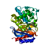
| ||||||||
|---|---|---|---|---|---|---|---|---|---|
| 1 |
| ||||||||
| Unit cell |
|
-Components
| #1: Protein | Mass: 31259.988 Da / Num. of mol.: 1 / Fragment: SVH-1 beta-lactamase Source method: isolated from a genetically manipulated source Source: (gene. exp.) Klebsiella pneumoniae (bacteria) / Gene: bla, shv1 / Production host: | ||||||||
|---|---|---|---|---|---|---|---|---|---|
| #2: Chemical | 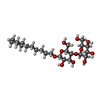  Mass: 508.600 Da / Num. of mol.: 2 / Source method: obtained synthetically / Formula: C24H44O11 Mass: 508.600 Da / Num. of mol.: 2 / Source method: obtained synthetically / Formula: C24H44O11#3: Chemical | ChemComp-1OG / |   Mass: 403.404 Da / Num. of mol.: 1 / Source method: obtained synthetically / Formula: C16H21NO9S Mass: 403.404 Da / Num. of mol.: 1 / Source method: obtained synthetically / Formula: C16H21NO9S#4: Chemical | ChemComp-EPE / |   Mass: 238.305 Da / Num. of mol.: 1 / Source method: obtained synthetically / Formula: C8H18N2O4S / Comment: pH buffer*YM Mass: 238.305 Da / Num. of mol.: 1 / Source method: obtained synthetically / Formula: C8H18N2O4S / Comment: pH buffer*YM#5: Water | ChemComp-HOH / |  Mass: 18.015 Da / Num. of mol.: 338 / Source method: isolated from a natural source / Formula: H2O Mass: 18.015 Da / Num. of mol.: 338 / Source method: isolated from a natural source / Formula: H2OHas protein modification | Y | |
-Experimental details
-Experiment
| Experiment | Method: X-RAY DIFFRACTION / Number of used crystals: 1 |
|---|
- Sample preparation
| Crystal | Density Matthews: 1.84 Å3/Da / Density % sol: 32.98 % |
|---|---|
| Crystal grow | Temperature: 293 K / Method: vapor diffusion, sitting drop Details: 21-30% PEG 6000, 0.1M HEPES buffer, 0.56mM Cymal-6, pH 6.8-7.8, VAPOR DIFFUSION, SITTING DROP, temperature 293K PH range: 6.8-7.8 |
-Data collection
| Diffraction | Mean temperature: 100 K |
|---|---|
| Diffraction source | Source: SYNCHROTRON / Site: SSRL  / Beamline: BL7-1 / Wavelength: 0.97946 Å / Beamline: BL7-1 / Wavelength: 0.97946 Å |
| Detector | Type: ADSC QUANTUM 315r / Detector: CCD / Date: Jul 1, 2010 |
| Radiation | Monochromator: Monochr. / Protocol: SINGLE WAVELENGTH / Monochromatic (M) / Laue (L): M / Scattering type: x-ray |
| Radiation wavelength | Wavelength: 0.97946 Å / Relative weight: 1 |
| Reflection | Resolution: 1.14→24.41 Å / Num. all: 79862 / Num. obs: 76013 / % possible obs: 95.18 % / Observed criterion σ(F): 1 / Observed criterion σ(I): 1 / Redundancy: 3.6 % / Rmerge(I) obs: 0.038 / Rsym value: 0.038 / Net I/σ(I): 13.8 |
| Reflection shell | Resolution: 1.14→1.18 Å / Redundancy: 3.2 % / Rsym value: 0.351 / % possible all: 77.2 |
- Processing
| Software |
| ||||||||||||||||||||||||||||||||||||||||||||||||||||||||||||||||||||||||||||||||||||||||||||||||||||||||||||||||||||||||||||||||||||||||||||||||||||||||||||||||||||||||||
|---|---|---|---|---|---|---|---|---|---|---|---|---|---|---|---|---|---|---|---|---|---|---|---|---|---|---|---|---|---|---|---|---|---|---|---|---|---|---|---|---|---|---|---|---|---|---|---|---|---|---|---|---|---|---|---|---|---|---|---|---|---|---|---|---|---|---|---|---|---|---|---|---|---|---|---|---|---|---|---|---|---|---|---|---|---|---|---|---|---|---|---|---|---|---|---|---|---|---|---|---|---|---|---|---|---|---|---|---|---|---|---|---|---|---|---|---|---|---|---|---|---|---|---|---|---|---|---|---|---|---|---|---|---|---|---|---|---|---|---|---|---|---|---|---|---|---|---|---|---|---|---|---|---|---|---|---|---|---|---|---|---|---|---|---|---|---|---|---|---|---|---|
| Refinement | Method to determine structure: MOLECULAR REPLACEMENT Starting model: pdb entry 1SHV Resolution: 1.14→24.41 Å / Cor.coef. Fo:Fc: 0.979 / Cor.coef. Fo:Fc free: 0.97 / SU B: 1.018 / SU ML: 0.022 / Cross valid method: THROUGHOUT / ESU R: 0.034 / ESU R Free: 0.035 / Stereochemistry target values: MAXIMUM LIKELIHOOD / Details: HYDROGENS HAVE BEEN ADDED IN THE RIDING POSITIONS
| ||||||||||||||||||||||||||||||||||||||||||||||||||||||||||||||||||||||||||||||||||||||||||||||||||||||||||||||||||||||||||||||||||||||||||||||||||||||||||||||||||||||||||
| Solvent computation | Ion probe radii: 0.8 Å / Shrinkage radii: 0.8 Å / VDW probe radii: 1.4 Å / Solvent model: MASK | ||||||||||||||||||||||||||||||||||||||||||||||||||||||||||||||||||||||||||||||||||||||||||||||||||||||||||||||||||||||||||||||||||||||||||||||||||||||||||||||||||||||||||
| Displacement parameters | Biso mean: 15.605 Å2
| ||||||||||||||||||||||||||||||||||||||||||||||||||||||||||||||||||||||||||||||||||||||||||||||||||||||||||||||||||||||||||||||||||||||||||||||||||||||||||||||||||||||||||
| Refinement step | Cycle: LAST / Resolution: 1.14→24.41 Å
| ||||||||||||||||||||||||||||||||||||||||||||||||||||||||||||||||||||||||||||||||||||||||||||||||||||||||||||||||||||||||||||||||||||||||||||||||||||||||||||||||||||||||||
| Refine LS restraints |
| ||||||||||||||||||||||||||||||||||||||||||||||||||||||||||||||||||||||||||||||||||||||||||||||||||||||||||||||||||||||||||||||||||||||||||||||||||||||||||||||||||||||||||
| LS refinement shell | Resolution: 1.142→1.172 Å / Total num. of bins used: 20
|
