 Movie
Movie Controller


+ Open data

- Basic information
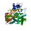
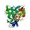
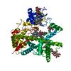
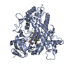
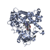
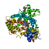
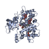
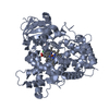
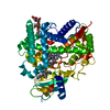
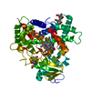
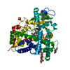
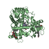
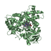
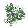
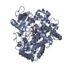
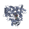
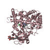
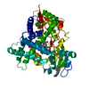
| Entry | Database: PDB / ID: 4jlt | ||||||
|---|---|---|---|---|---|---|---|
| Title | Crystal structure of P450 2B4(H226Y) in complex with paroxetine | ||||||
 Components Components | Cytochrome P450 2B4 | ||||||
 Keywords Keywords | OXIDOREDUCTASE / P450 / cytochrome P450 2B4 / monooxygenase / ME protein / CYP 2B4 | ||||||
| Function / homology |  Function and homology information Function and homology informationarachidonate epoxygenase activity / epoxygenase P450 pathway / oxidoreductase activity, acting on paired donors, with incorporation or reduction of molecular oxygen, reduced flavin or flavoprotein as one donor, and incorporation of one atom of oxygen / unspecific monooxygenase / xenobiotic metabolic process / iron ion binding / heme binding / endoplasmic reticulum membrane Similarity search - Function | ||||||
| Biological species |  | ||||||
| Method |  X-RAY DIFFRACTION / SYNCHROTRON / MOLECULAR REPLACEMENT / Resolution: 2.14 Å X-RAY DIFFRACTION / SYNCHROTRON / MOLECULAR REPLACEMENT / Resolution: 2.14 Å | ||||||
 Authors Authors | Shah, M.B. / Pascual, J. / Stout, C.D. / Halpert, J.R. | ||||||
 Citation Citation | Journal: J.Pharmacol.Exp.Ther. / Year: 2013 Title: A Structural Snapshot of CYP2B4 in Complex with Paroxetine Provides Insights into Ligand Binding and Clusters of Conformational States. Authors: Shah, M.B. / Kufareva, I. / Pascual, J. / Zhang, Q. / Stout, C.D. / Halpert, J.R. | ||||||
| History |
|
- Structure visualization
| Structure viewer | Molecule: MolmilJmol/JSmol |
|---|
- Downloads & links
-Download
| PDBx/mmCIF format | 4jlt.cif.gz | 118.8 KB | Display | PDBx/mmCIF format |
|---|---|---|---|---|
| PDB format | pdb4jlt.ent.gz | 88.4 KB | Display | PDB format |
| PDBx/mmJSON format | 4jlt.json.gz | Tree view | PDBx/mmJSON format | |
| Others |  Other downloads Other downloads |
-Validation report
| Arichive directory | https://data.pdbj.org/pub/pdb/validation_reports/jl/4jltftp://data.pdbj.org/pub/pdb/validation_reports/jl/4jlt | HTTPS FTP |
|---|
-Related structure data
| Related structure data | 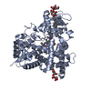 3mvrS S: Starting model for refinement |
|---|---|
| Similar structure data |


-Links
 PDBj
PDBj


- Assembly
| Deposited unit | 
| ||||||||
|---|---|---|---|---|---|---|---|---|---|
| 1 |
| ||||||||
| Unit cell |
|
-Components
-Protein , 1 types, 1 molecules A
| #1: Protein | Mass: 54169.082 Da / Num. of mol.: 1 / Fragment: Cytochrome P450 2B4 / Mutation: H226Y Source method: isolated from a genetically manipulated source Source: (gene. exp.)  |
|---|
-Non-polymers , 7 types, 231 molecules 


| #2: Chemical | ChemComp-HEM /  Mass: 616.487 Da / Num. of mol.: 1 / Source method: obtained synthetically / Formula: C34H32FeN4O4 Mass: 616.487 Da / Num. of mol.: 1 / Source method: obtained synthetically / Formula: C34H32FeN4O4 | ||||||||||
|---|---|---|---|---|---|---|---|---|---|---|---|
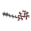
| #3: Chemical |  Mass: 494.573 Da / Num. of mol.: 3 / Source method: obtained synthetically / Formula: C23H42O11 / Comment: detergent*YM Mass: 494.573 Da / Num. of mol.: 3 / Source method: obtained synthetically / Formula: C23H42O11 / Comment: detergent*YM#4: Chemical | ChemComp-8PR / |  Mass: 329.365 Da / Num. of mol.: 1 / Source method: obtained synthetically / Formula: C19H20FNO3 / Comment: medication, antidepressant, inhibitor*YM Mass: 329.365 Da / Num. of mol.: 1 / Source method: obtained synthetically / Formula: C19H20FNO3 / Comment: medication, antidepressant, inhibitor*YM#5: Chemical | ChemComp-SO4 /  Mass: 96.063 Da / Num. of mol.: 4 / Source method: obtained synthetically / Formula: SO4 Mass: 96.063 Da / Num. of mol.: 4 / Source method: obtained synthetically / Formula: SO4#6: Chemical | ChemComp-GOL / |  Mass: 92.094 Da / Num. of mol.: 1 / Source method: obtained synthetically / Formula: C3H8O3 Mass: 92.094 Da / Num. of mol.: 1 / Source method: obtained synthetically / Formula: C3H8O3#7: Chemical |  Mass: 94.971 Da / Num. of mol.: 2 / Source method: obtained synthetically / Formula: PO4 Mass: 94.971 Da / Num. of mol.: 2 / Source method: obtained synthetically / Formula: PO4#8: Water | ChemComp-HOH / | Mass: 18.015 Da / Num. of mol.: 219 / Source method: isolated from a natural source / Formula: H2O |
-Details
| Sequence details | AUTHORS INFORMED THAT THE GENBANK SEQUENCE IS THOUGHT TO CONTAIN A SEQUENCING |
|---|
-Experimental details
-Experiment
| Experiment | Method: X-RAY DIFFRACTION / Number of used crystals: 1 |
|---|
- Sample preparation
| Crystal | Density Matthews: 3.37 Å3/Da / Density % sol: 63.54 % |
|---|---|
| Crystal grow | Temperature: 291 K / Method: vapor diffusion, sitting drop / pH: 7.5 Details: 0.1 M HEPES, 2M Ammonium Sulfate, pH 7.5, VAPOR DIFFUSION, SITTING DROP, temperature 291K |
-Data collection
| Diffraction | Mean temperature: 100 K |
|---|---|
| Diffraction source | Source: SYNCHROTRON / Site: SSRL  / Beamline: BL11-1 / Wavelength: 0.98 / Wavelength: 0.98 Å / Beamline: BL11-1 / Wavelength: 0.98 / Wavelength: 0.98 Å |
| Detector | Type: MARMOSAIC 325 mm CCD / Detector: CCD / Date: Dec 11, 2010 |
| Radiation | Monochromator: SI(111) / Protocol: SINGLE WAVELENGTH / Monochromatic (M) / Laue (L): M / Scattering type: x-ray |
| Radiation wavelength | Wavelength: 0.98 Å / Relative weight: 1 |
| Reflection | Resolution: 2.137→78.6 Å / Num. all: 41220 / Num. obs: 41055 / % possible obs: 99.6 % / Observed criterion σ(F): 0 / Observed criterion σ(I): 0 / Redundancy: 11.1 % / Rmerge(I) obs: 0.064 / Rsym value: 0.064 / Net I/σ(I): 8.2 |
| Reflection shell | Resolution: 2.137→2.25 Å / Redundancy: 11.1 % / Rmerge(I) obs: 0.444 / Mean I/σ(I) obs: 1.7 / Rsym value: 0.444 / % possible all: 99.4 |
- Processing
| Software |
| ||||||||||||||||||||||||||||||||||||||||||||||||||||||||||||||||||||||||||||||||||||||||||||||||||||||||||||||||||||||||||||||||||||||||||||||||||||||||||||||||||||||||||
|---|---|---|---|---|---|---|---|---|---|---|---|---|---|---|---|---|---|---|---|---|---|---|---|---|---|---|---|---|---|---|---|---|---|---|---|---|---|---|---|---|---|---|---|---|---|---|---|---|---|---|---|---|---|---|---|---|---|---|---|---|---|---|---|---|---|---|---|---|---|---|---|---|---|---|---|---|---|---|---|---|---|---|---|---|---|---|---|---|---|---|---|---|---|---|---|---|---|---|---|---|---|---|---|---|---|---|---|---|---|---|---|---|---|---|---|---|---|---|---|---|---|---|---|---|---|---|---|---|---|---|---|---|---|---|---|---|---|---|---|---|---|---|---|---|---|---|---|---|---|---|---|---|---|---|---|---|---|---|---|---|---|---|---|---|---|---|---|---|---|---|---|
| Refinement | Method to determine structure: MOLECULAR REPLACEMENT Starting model: PDB ENTRY 3MVR Resolution: 2.14→78.6 Å / Cor.coef. Fo:Fc: 0.958 / Cor.coef. Fo:Fc free: 0.939 / Occupancy max: 1 / Occupancy min: 1 / SU B: 4.243 / SU ML: 0.112 / Cross valid method: THROUGHOUT / ESU R: 0.172 / ESU R Free: 0.161 / Stereochemistry target values: MAXIMUM LIKELIHOOD / Details: HYDROGENS HAVE BEEN ADDED IN THE RIDING POSITIONS
| ||||||||||||||||||||||||||||||||||||||||||||||||||||||||||||||||||||||||||||||||||||||||||||||||||||||||||||||||||||||||||||||||||||||||||||||||||||||||||||||||||||||||||
| Solvent computation | Ion probe radii: 0.8 Å / Shrinkage radii: 0.8 Å / VDW probe radii: 1.4 Å / Solvent model: MASK | ||||||||||||||||||||||||||||||||||||||||||||||||||||||||||||||||||||||||||||||||||||||||||||||||||||||||||||||||||||||||||||||||||||||||||||||||||||||||||||||||||||||||||
| Displacement parameters | Biso max: 172.23 Å2 / Biso mean: 40.305 Å2 / Biso min: 15.74 Å2
| ||||||||||||||||||||||||||||||||||||||||||||||||||||||||||||||||||||||||||||||||||||||||||||||||||||||||||||||||||||||||||||||||||||||||||||||||||||||||||||||||||||||||||
| Refinement step | Cycle: LAST / Resolution: 2.14→78.6 Å
| ||||||||||||||||||||||||||||||||||||||||||||||||||||||||||||||||||||||||||||||||||||||||||||||||||||||||||||||||||||||||||||||||||||||||||||||||||||||||||||||||||||||||||
| Refine LS restraints |
| ||||||||||||||||||||||||||||||||||||||||||||||||||||||||||||||||||||||||||||||||||||||||||||||||||||||||||||||||||||||||||||||||||||||||||||||||||||||||||||||||||||||||||
| LS refinement shell | Resolution: 2.14→2.19 Å / Total num. of bins used: 20
|
