 Movie
Movie Controller

[English] 日本語
 Yorodumi
- PDB-4jhw: Crystal Structure of Respiratory Syncytial Virus Fusion Glycoprot... -
+ Open data

- Basic information
| Entry | Database: PDB / ID: 4jhw | ||||||
|---|---|---|---|---|---|---|---|
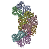
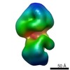
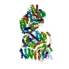
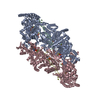
| Title | Crystal Structure of Respiratory Syncytial Virus Fusion Glycoprotein Stabilized in the Prefusion Conformation by Human Antibody D25 | ||||||
 Components Components |
| ||||||
 Keywords Keywords | IMMUNE SYSTEM / immunoglobulin / type I fusion protein / membrane fusion | ||||||
| Function / homology |  Function and homology information Function and homology informationsymbiont-mediated induction of syncytium formation / Translation of respiratory syncytial virus mRNAs / RSV-host interactions / Assembly and release of respiratory syncytial virus (RSV) virions / Maturation of hRSV A proteins / Respiratory syncytial virus (RSV) attachment and entry / host cell Golgi membrane / entry receptor-mediated virion attachment to host cell / fusion of virus membrane with host plasma membrane / viral envelope ...symbiont-mediated induction of syncytium formation / Translation of respiratory syncytial virus mRNAs / RSV-host interactions / Assembly and release of respiratory syncytial virus (RSV) virions / Maturation of hRSV A proteins / Respiratory syncytial virus (RSV) attachment and entry / host cell Golgi membrane / entry receptor-mediated virion attachment to host cell / fusion of virus membrane with host plasma membrane / viral envelope / symbiont entry into host cell / host cell plasma membrane / virion membrane / identical protein binding / plasma membrane Similarity search - Function | ||||||
| Biological species |  Homo sapiens (human) Homo sapiens (human) Human respiratory syncytial virus Human respiratory syncytial virus | ||||||
| Method |  X-RAY DIFFRACTION / SYNCHROTRON / MOLECULAR REPLACEMENT / Resolution: 3.6 Å X-RAY DIFFRACTION / SYNCHROTRON / MOLECULAR REPLACEMENT / Resolution: 3.6 Å | ||||||
 Authors Authors | Mclellan, J.S. / Chen, M. / Leung, S. / Graepel, K.W. / Du, X. / Yang, Y. / Zhou, T. / Baxa, U. / Yasuda, E. / Beaumont, T. ...Mclellan, J.S. / Chen, M. / Leung, S. / Graepel, K.W. / Du, X. / Yang, Y. / Zhou, T. / Baxa, U. / Yasuda, E. / Beaumont, T. / Kumar, A. / Modjarrad, K. / Zheng, Z. / Zhao, M. / Xia, N. / Kwong, P.D. / Graham, B.S. | ||||||
 Citation Citation | Journal: Science / Year: 2013 Title: Structure of RSV fusion glycoprotein trimer bound to a prefusion-specific neutralizing antibody. Authors: McLellan, J.S. / Chen, M. / Leung, S. / Graepel, K.W. / Du, X. / Yang, Y. / Zhou, T. / Baxa, U. / Yasuda, E. / Beaumont, T. / Kumar, A. / Modjarrad, K. / Zheng, Z. / Zhao, M. / Xia, N. / ...Authors: McLellan, J.S. / Chen, M. / Leung, S. / Graepel, K.W. / Du, X. / Yang, Y. / Zhou, T. / Baxa, U. / Yasuda, E. / Beaumont, T. / Kumar, A. / Modjarrad, K. / Zheng, Z. / Zhao, M. / Xia, N. / Kwong, P.D. / Graham, B.S. | ||||||
| History |
|
- Structure visualization
| Structure viewer | Molecule: MolmilJmol/JSmol |
|---|
- Downloads & links
-Download
| PDBx/mmCIF format | 4jhw.cif.gz | 364.8 KB | Display | PDBx/mmCIF format |
|---|---|---|---|---|
| PDB format | pdb4jhw.ent.gz | 300.9 KB | Display | PDB format |
| PDBx/mmJSON format | 4jhw.json.gz | Tree view | PDBx/mmJSON format | |
| Others |  Other downloads Other downloads |
-Validation report
| Arichive directory | https://data.pdbj.org/pub/pdb/validation_reports/jh/4jhwftp://data.pdbj.org/pub/pdb/validation_reports/jh/4jhw | HTTPS FTP |
|---|
-Related structure data
| Related structure data |  4jhaSC  3rrrS S: Starting model for refinement C: citing same article ( |
|---|---|
| Similar structure data |

-Links
 PDBj
PDBj


- Assembly
| Deposited unit | 
| ||||||||
|---|---|---|---|---|---|---|---|---|---|
| 1 | 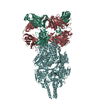
| ||||||||
| Unit cell |
|
-Components
| #1: Antibody | Mass: 24375.449 Da / Num. of mol.: 1 Source method: isolated from a genetically manipulated source Source: (gene. exp.) Homo sapiens (human) / Plasmid: pVRC8400 / Cell line (production host): HEK293 / Production host: homo sapiens (human) |
|---|---|
| #2: Antibody | Mass: 23325.865 Da / Num. of mol.: 1 Source method: isolated from a genetically manipulated source Source: (gene. exp.) Homo sapiens (human) / Plasmid: pVRC8400 / Cell line (production host): HEK293 / Production host: homo sapiens (human) |
| #3: Protein | Mass: 55014.629 Da / Num. of mol.: 1 / Mutation: P102A, I379V, M447V Source method: isolated from a genetically manipulated source Source: (gene. exp.) Human respiratory syncytial virus / Strain: A2 / Gene: F, fusion glycoprotein / Plasmid: paH / Cell line (production host): HEK293 / Production host: homo sapiens (human) / References: UniProt: P03420 |
| Has protein modification | Y |
| Sequence details | AUTHOR STATES THAT THE ORF FOR THE CRYSTALLIZED RSV F PROTEIN IS: ...AUTHOR STATES THAT THE ORF FOR THE CRYSTALLIZ |
-Experimental details
-Experiment
| Experiment | Method: X-RAY DIFFRACTION / Number of used crystals: 1 |
|---|
- Sample preparation
| Crystal | Density Matthews: 2.87 Å3/Da / Density % sol: 57.07 % |
|---|---|
| Crystal grow | Temperature: 293 K / Method: vapor diffusion, hanging drop / pH: 7.5 Details: 30% (w/v) PEG 400, 3.75% (w/v) PEG 3350, 0.1 M HEPES pH 7.5, and 1% (v/v) 1,2-butanediol, VAPOR DIFFUSION, HANGING DROP, temperature 293K |
-Data collection
| Diffraction | Mean temperature: 200 K |
|---|---|
| Diffraction source | Source: SYNCHROTRON / Site: APS  / Beamline: 22-ID / Wavelength: 1 Å / Beamline: 22-ID / Wavelength: 1 Å |
| Detector | Type: MARMOSAIC 300 mm CCD / Detector: CCD / Date: Dec 19, 2012 |
| Radiation | Monochromator: Si 111. Rosenbaum-Rock double-crystal monochromator Protocol: SINGLE WAVELENGTH / Monochromatic (M) / Laue (L): M / Scattering type: x-ray |
| Radiation wavelength | Wavelength: 1 Å / Relative weight: 1 |
| Reflection | Resolution: 3.6→50 Å / Num. all: 13953 / Num. obs: 13898 / % possible obs: 99.6 % / Observed criterion σ(F): 1 / Observed criterion σ(I): 1 |
| Reflection shell | Resolution: 3.6→3.73 Å / Redundancy: 5.2 % / % possible all: 99.3 |
- Processing
| Software |
| ||||||||||||||||||||||||||||||||||||||||||||||||||||||||||||||||||||||||||||||||||||||||||||||||||||||||||||||||||||||||||||||||||||||||||||||||||||||||||||||||||||||||||||||||||||||||||||||||||||||||||||||||||||||||||||||||||||||||||||||||||||||||||
|---|---|---|---|---|---|---|---|---|---|---|---|---|---|---|---|---|---|---|---|---|---|---|---|---|---|---|---|---|---|---|---|---|---|---|---|---|---|---|---|---|---|---|---|---|---|---|---|---|---|---|---|---|---|---|---|---|---|---|---|---|---|---|---|---|---|---|---|---|---|---|---|---|---|---|---|---|---|---|---|---|---|---|---|---|---|---|---|---|---|---|---|---|---|---|---|---|---|---|---|---|---|---|---|---|---|---|---|---|---|---|---|---|---|---|---|---|---|---|---|---|---|---|---|---|---|---|---|---|---|---|---|---|---|---|---|---|---|---|---|---|---|---|---|---|---|---|---|---|---|---|---|---|---|---|---|---|---|---|---|---|---|---|---|---|---|---|---|---|---|---|---|---|---|---|---|---|---|---|---|---|---|---|---|---|---|---|---|---|---|---|---|---|---|---|---|---|---|---|---|---|---|---|---|---|---|---|---|---|---|---|---|---|---|---|---|---|---|---|---|---|---|---|---|---|---|---|---|---|---|---|---|---|---|---|---|---|---|---|---|---|---|---|---|---|---|---|---|---|---|---|---|
| Refinement | Method to determine structure: MOLECULAR REPLACEMENT Starting model: 3RRR AND 4JHA Resolution: 3.6→42.237 Å / SU ML: 0.51 / σ(F): 1.35 / Phase error: 32.41 / Stereochemistry target values: ML
| ||||||||||||||||||||||||||||||||||||||||||||||||||||||||||||||||||||||||||||||||||||||||||||||||||||||||||||||||||||||||||||||||||||||||||||||||||||||||||||||||||||||||||||||||||||||||||||||||||||||||||||||||||||||||||||||||||||||||||||||||||||||||||
| Solvent computation | Shrinkage radii: 0.9 Å / VDW probe radii: 1.11 Å / Solvent model: FLAT BULK SOLVENT MODEL | ||||||||||||||||||||||||||||||||||||||||||||||||||||||||||||||||||||||||||||||||||||||||||||||||||||||||||||||||||||||||||||||||||||||||||||||||||||||||||||||||||||||||||||||||||||||||||||||||||||||||||||||||||||||||||||||||||||||||||||||||||||||||||
| Refinement step | Cycle: LAST / Resolution: 3.6→42.237 Å
| ||||||||||||||||||||||||||||||||||||||||||||||||||||||||||||||||||||||||||||||||||||||||||||||||||||||||||||||||||||||||||||||||||||||||||||||||||||||||||||||||||||||||||||||||||||||||||||||||||||||||||||||||||||||||||||||||||||||||||||||||||||||||||
| Refine LS restraints |
| ||||||||||||||||||||||||||||||||||||||||||||||||||||||||||||||||||||||||||||||||||||||||||||||||||||||||||||||||||||||||||||||||||||||||||||||||||||||||||||||||||||||||||||||||||||||||||||||||||||||||||||||||||||||||||||||||||||||||||||||||||||||||||
| LS refinement shell |
| ||||||||||||||||||||||||||||||||||||||||||||||||||||||||||||||||||||||||||||||||||||||||||||||||||||||||||||||||||||||||||||||||||||||||||||||||||||||||||||||||||||||||||||||||||||||||||||||||||||||||||||||||||||||||||||||||||||||||||||||||||||||||||
| Refinement TLS params. | Method: refined / Refine-ID: X-RAY DIFFRACTION
| ||||||||||||||||||||||||||||||||||||||||||||||||||||||||||||||||||||||||||||||||||||||||||||||||||||||||||||||||||||||||||||||||||||||||||||||||||||||||||||||||||||||||||||||||||||||||||||||||||||||||||||||||||||||||||||||||||||||||||||||||||||||||||
| Refinement TLS group |
|
