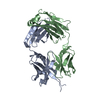
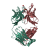
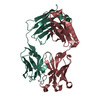
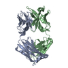
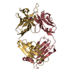
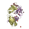
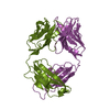
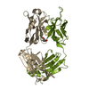
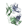
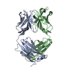
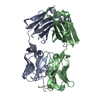
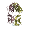
- PDB-4j8r: Structure of an octapeptide repeat of the prion protein bound to ... -
+
Open data
ID or keywords:
Loading...
-
Basic information
Entry
Database: PDB / ID: 4j8r
Title
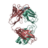
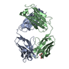
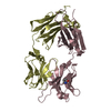
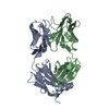
Structure of an octapeptide repeat of the prion protein bound to the POM2 Fab antibody fragment
Components
Heavy chain of POM2 Fab
Light chain of POM2 Fab
Major prion protein
Keywords
IMMUNE SYSTEM / Immunoglobulin fold / Fab / Antibody / Octapeptide repeat / Mouse prion protein
Function / homology
Function and homology information
Insertion of tail-anchored proteins into the endoplasmic reticulum membrane / negative regulation of amyloid precursor protein catabolic process / regulation of glutamate receptor signaling pathway / lamin binding / aspartic-type endopeptidase inhibitor activity / regulation of calcium ion import across plasma membrane / glycosaminoglycan binding / regulation of potassium ion transmembrane transport / positive regulation of glutamate receptor signaling pathway / cupric ion binding ...Insertion of tail-anchored proteins into the endoplasmic reticulum membrane / negative regulation of amyloid precursor protein catabolic process / regulation of glutamate receptor signaling pathway / lamin binding / aspartic-type endopeptidase inhibitor activity / regulation of calcium ion import across plasma membrane / glycosaminoglycan binding / regulation of potassium ion transmembrane transport / positive regulation of glutamate receptor signaling pathway / cupric ion binding / negative regulation of interleukin-17 production / ATP-dependent protein binding / type 5 metabotropic glutamate receptor binding / nucleobase-containing compound metabolic process / negative regulation of dendritic spine maintenance / negative regulation of calcineurin-NFAT signaling cascade / negative regulation of interleukin-2 production / response to copper ion / negative regulation of activated T cell proliferation / negative regulation of amyloid-beta formation / response to amyloid-beta / negative regulation of type II interferon production / cuprous ion binding / negative regulation of long-term synaptic potentiation / intracellular copper ion homeostasis / positive regulation of protein targeting to membrane / negative regulation of T cell receptor signaling pathway / response to cadmium ion / side of membrane / positive regulation of calcium-mediated signaling / neuron projection maintenance / inclusion body / cellular response to copper ion / molecular function activator activity / positive regulation of protein localization to plasma membrane / molecular condensate scaffold activity / tubulin binding / cellular response to xenobiotic stimulus / protein homooligomerization / protein destabilization / cellular response to amyloid-beta / terminal bouton / positive regulation of neuron apoptotic process / regulation of protein localization / amyloid-beta binding / protein-folding chaperone binding / signaling receptor activity / protease binding / response to oxidative stress / nuclear membrane / microtubule binding / molecular adaptor activity / transmembrane transporter binding / learning or memory / mitochondrial outer membrane / postsynaptic density / intracellular signal transduction / membrane raft / copper ion binding / dendrite / negative regulation of apoptotic process / protein-containing complex binding / Golgi apparatus / negative regulation of transcription by RNA polymerase II / cell surface / endoplasmic reticulum / membrane / metal ion binding / identical protein binding / plasma membrane / cytosol Similarity search - Function
Prion, copper binding octapeptide repeat / Copper binding octapeptide repeat region / Major prion protein N-terminal domain / Major prion protein bPrPp - N terminal / Prion protein signature 1. / Prion protein signature 2. / Prion protein / Major prion protein / Prion/Doppel protein, beta-ribbon domain / Prion/Doppel beta-ribbon domain superfamily ...Prion, copper binding octapeptide repeat / Copper binding octapeptide repeat region / Major prion protein N-terminal domain / Major prion protein bPrPp - N terminal / Prion protein signature 1. / Prion protein signature 2. / Prion protein / Major prion protein / Prion/Doppel protein, beta-ribbon domain / Prion/Doppel beta-ribbon domain superfamily / Prion/Doppel alpha-helical domain / Immunoglobulins / Immunoglobulin-like / Sandwich / Mainly Beta Similarity search - Domain/homology
A: Light chain of POM2 Fab B: Heavy chain of POM2 Fab I: Major prion protein C: Light chain of POM2 Fab D: Heavy chain of POM2 Fab J: Major prion protein
Resolution: 2.303→39.597 Å / SU ML: 0.33 / σ(F): 1.34 / Phase error: 27.31 / Stereochemistry target values: ML Details: The Rmeas for the data collection is 0.161 (overall data)
Rfactor
Num. reflection
% reflection
Rfree
0.2574
2215
5.03 %
Rwork
0.2468
-
-
obs
0.2474
44006
99.83 %
all
-
41792
-
Solvent computation
Shrinkage radii: 0.9 Å / VDW probe radii: 1.11 Å / Solvent model: FLAT BULK SOLVENT MODEL
Refine analyze
Luzzati coordinate error obs: 0.39 Å / Luzzati d res low obs: 2.3 Å
Refinement step
Cycle: LAST / Resolution: 2.303→39.597 Å
Protein
Nucleic acid
Ligand
Solvent
Total
Num. atoms
6626
0
0
165
6791
Refine LS restraints
Refine-ID
Type
Dev ideal
Number
X-RAY DIFFRACTION
f_bond_d
0.028
6797
X-RAY DIFFRACTION
f_angle_d
1.933
9247
X-RAY DIFFRACTION
f_dihedral_angle_d
15.78
2403
X-RAY DIFFRACTION
f_chiral_restr
0.167
1040
X-RAY DIFFRACTION
f_plane_restr
0.013
1179
LS refinement shell
Resolution (Å)
Rfactor Rfree
Num. reflection Rfree
Rfactor Rwork
Num. reflection Rwork
Refine-ID
% reflection obs (%)
2.3029-2.353
0.3471
138
0.3406
2504
X-RAY DIFFRACTION
98
2.353-2.4077
0.3209
137
0.3498
2592
X-RAY DIFFRACTION
100
2.4077-2.4679
0.3955
138
0.3401
2569
X-RAY DIFFRACTION
100
2.4679-2.5346
0.3402
132
0.3287
2574
X-RAY DIFFRACTION
100
2.5346-2.6092
0.3446
130
0.3169
2600
X-RAY DIFFRACTION
100
2.6092-2.6934
0.3035
140
0.3172
2567
X-RAY DIFFRACTION
100
2.6934-2.7897
0.3293
146
0.3104
2564
X-RAY DIFFRACTION
100
2.7897-2.9013
0.3204
124
0.2982
2605
X-RAY DIFFRACTION
100
2.9013-3.0333
0.3067
130
0.2808
2623
X-RAY DIFFRACTION
100
3.0333-3.1932
0.3208
149
0.2743
2578
X-RAY DIFFRACTION
100
3.1932-3.3931
0.2991
161
0.2567
2590
X-RAY DIFFRACTION
100
3.3931-3.655
0.2548
134
0.2383
2625
X-RAY DIFFRACTION
100
3.655-4.0224
0.2266
150
0.2184
2625
X-RAY DIFFRACTION
100
4.0224-4.6037
0.1686
131
0.1912
2650
X-RAY DIFFRACTION
100
4.6037-5.7973
0.1984
138
0.2
2684
X-RAY DIFFRACTION
100
5.7973-39.6031
0.2358
137
0.2326
2841
X-RAY DIFFRACTION
100
Refinement TLS params.
Method: refined / Origin x: 11.0193 Å / Origin y: -13.508 Å / Origin z: 27.3858 Å
11
12
13
21
22
23
31
32
33
T
0.3912 Å2
-0.0266 Å2
-0.037 Å2
-
0.3517 Å2
0.0073 Å2
-
-
0.4468 Å2
L
-0.1498 °2
-0.1973 °2
0.0299 °2
-
0.3556 °2
-0.1949 °2
-
-
0.0682 °2
S
-0.0202 Å °
0.0084 Å °
-0.0082 Å °
-0.0262 Å °
0.021 Å °
0.0284 Å °
-0.0098 Å °
-0.0005 Å °
-0.0011 Å °
Refinement TLS group
Selection details: all
+
About Yorodumi
-
News
-
Feb 9, 2022. New format data for meta-information of EMDB entries
New format data for meta-information of EMDB entries
Version 3 of the EMDB header file is now the official format.
The previous official version 1.9 will be removed from the archive.
In the structure databanks used in Yorodumi, some data are registered as the other names, "COVID-19 virus" and "2019-nCoV". Here are the details of the virus and the list of structure data.
Jan 31, 2019. EMDB accession codes are about to change! (news from PDBe EMDB page)
EMDB accession codes are about to change! (news from PDBe EMDB page)
The allocation of 4 digits for EMDB accession codes will soon come to an end. Whilst these codes will remain in use, new EMDB accession codes will include an additional digit and will expand incrementally as the available range of codes is exhausted. The current 4-digit format prefixed with “EMD-” (i.e. EMD-XXXX) will advance to a 5-digit format (i.e. EMD-XXXXX), and so on. It is currently estimated that the 4-digit codes will be depleted around Spring 2019, at which point the 5-digit format will come into force.
The EM Navigator/Yorodumi systems omit the EMD- prefix.
Related info.:Q: What is EMD? / ID/Accession-code notation in Yorodumi/EM Navigator
Yorodumi is a browser for structure data from EMDB, PDB, SASBDB, etc.
This page is also the successor to EM Navigator detail page, and also detail information page/front-end page for Omokage search.
The word "yorodu" (or yorozu) is an old Japanese word meaning "ten thousand". "mi" (miru) is to see.
Related info.:EMDB / PDB / SASBDB / Comparison of 3 databanks / Yorodumi Search / Aug 31, 2016. New EM Navigator & Yorodumi / Yorodumi Papers / Jmol/JSmol / Function and homology information / Changes in new EM Navigator and Yorodumi
 Movie
Movie Controller

 Yorodumi
 Open data
 Basic information
 Components
 Keywords
 Function and homology information
 X-RAY DIFFRACTION /
  Authors
 Citation
 Structure visualization
 Downloads & links
 Other downloads


 PDBj
PDBj


 Assembly
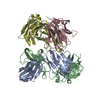
 Mass: 18.015 Da / Num. of mol.: 165 / Source method: isolated from a natural source / Formula: H2O
 Sample preparation
 / Beamline: BL9-2 / Wavelength: 0.979 Å
 Processing