[English] 日本語
Yorodumi
- PDB-4hss: Structure of the Full-Length Major Pilin SpaD from Corynebacteriu... -

+
Open data


ID or keywords:

Loading...

-
Basic information

Entry
Database: PDB / ID: 4hss
TitleStructure of the Full-Length Major Pilin SpaD from Corynebacterium diphtheriae
ComponentsPutative fimbrial subunit
KeywordsCELL ADHESION / CnaA/CnaB folded domains / Major pilin polymer / Homopolymer / Isopeptide bonding between Lys and Asn sidechains
Function / homology
Function and homology information


carbohydrate metabolic process / membrane / metal ion binding
Similarity search - Function
Sgo0707-like, N2 domain / Sgo0707 N2 domain / : / Fimbrial isopeptide formation D2 domain / Immunoglobulin-like - #740 / Prealbumin-like fold domain / Prealbumin-like fold domain / Immunoglobulins / Immunoglobulin-like fold / Immunoglobulin-like ...Sgo0707-like, N2 domain / Sgo0707 N2 domain / : / Fimbrial isopeptide formation D2 domain / Immunoglobulin-like - #740 / Prealbumin-like fold domain / Prealbumin-like fold domain / Immunoglobulins / Immunoglobulin-like fold / Immunoglobulin-like / Sandwich / Mainly Beta
Similarity search - Domain/homology
Biological speciesCorynebacterium diphtheriae (bacteria)
MethodX-RAY DIFFRACTION / SYNCHROTRON / MOLECULAR REPLACEMENT / Resolution: 2.5 Å
AuthorsPaterson, N.G. / Kang, H.J. / Baker, E.N.
CitationJournal: Acta Crystallogr.,Sect.D / Year: 2014
Title: A slow-forming isopeptide bond in the structure of the major pilin SpaD from Corynebacterium diphtheriae has implications for pilus assembly.
Authors: Kang, H.J. / Paterson, N.G. / Kim, C.U. / Middleditch, M. / Chang, C. / Ton-That, H. / Baker, E.N.
History
DepositionOct 30, 2012Deposition site: RCSB / Processing site: RCSB
Revision 1.0Nov 13, 2013Provider: repository / Type: Initial release
Revision 1.1May 14, 2014Group: Database references
Revision 1.2Jun 25, 2014Group: Database references
Revision 1.3Sep 20, 2023Group: Data collection / Database references ...Data collection / Database references / Derived calculations / Refinement description
Category: chem_comp_atom / chem_comp_bond ...chem_comp_atom / chem_comp_bond / database_2 / pdbx_initial_refinement_model / pdbx_struct_conn_angle / struct_conn / struct_ref_seq_dif / struct_site
Item: _database_2.pdbx_DOI / _database_2.pdbx_database_accession ..._database_2.pdbx_DOI / _database_2.pdbx_database_accession / _pdbx_struct_conn_angle.ptnr1_auth_comp_id / _pdbx_struct_conn_angle.ptnr1_auth_seq_id / _pdbx_struct_conn_angle.ptnr1_label_comp_id / _pdbx_struct_conn_angle.ptnr1_label_seq_id / _pdbx_struct_conn_angle.ptnr3_auth_comp_id / _pdbx_struct_conn_angle.ptnr3_auth_seq_id / _pdbx_struct_conn_angle.ptnr3_label_comp_id / _pdbx_struct_conn_angle.ptnr3_label_seq_id / _pdbx_struct_conn_angle.value / _struct_conn.pdbx_dist_value / _struct_conn.pdbx_leaving_atom_flag / _struct_conn.ptnr1_auth_asym_id / _struct_conn.ptnr1_auth_comp_id / _struct_conn.ptnr1_auth_seq_id / _struct_conn.ptnr1_label_asym_id / _struct_conn.ptnr1_label_comp_id / _struct_conn.ptnr1_label_seq_id / _struct_conn.ptnr2_auth_asym_id / _struct_conn.ptnr2_auth_seq_id / _struct_conn.ptnr2_label_asym_id / _struct_conn.ptnr2_label_seq_id / _struct_ref_seq_dif.details / _struct_site.pdbx_auth_asym_id / _struct_site.pdbx_auth_comp_id / _struct_site.pdbx_auth_seq_id

-
Structure visualization

Structure viewerMolecule:
MolmilJmol/JSmol

Downloads & links

-
Assembly

Deposited unit
A: Putative fimbrial subunit
B: Putative fimbrial subunit
hetero molecules


Theoretical massNumber of molelcules
Total (without water)94,3234
Polymers94,2432
Non-polymers802
Water5,044280
1
A: Putative fimbrial subunit
hetero molecules


Theoretical massNumber of molelcules
Total (without water)47,1622
Polymers47,1221
Non-polymers401
Water181
TypeNameSymmetry operationNumber
identity operation1_555x,y,z1
MethodPISA
2
B: Putative fimbrial subunit
hetero molecules


Theoretical massNumber of molelcules
Total (without water)47,1622
Polymers47,1221
Non-polymers401
Water181
TypeNameSymmetry operationNumber
identity operation1_555x,y,z1
MethodPISA
Unit cell
Length a, b, c (Å)32.828, 56.791, 435.839
Angle α, β, γ (deg.)90.00, 90.00, 90.00
Int Tables number19
Space group name H-MP212121
Details
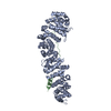
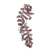
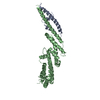
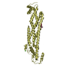
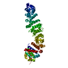
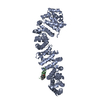
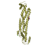
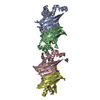
SpaD subunits covalently linked by sortase-catalysed isopeptide bond between C-terminal sortase motif Thr carboxyl and epsilon amino group of Lys 179 in D1 of next monomer. Chains A and B are arranged in the predicted polymer orientation and this continues throughout the crystal via symmetry operations.

-
Components

#1: Protein Putative fimbrial subunit


Mass: 47121.578 Da / Num. of mol.: 2 / Fragment: Domains D1-D3
Source method: isolated from a genetically manipulated source
Source: (gene. exp.) Corynebacterium diphtheriae (bacteria) / Strain: ATCC 700971 / NCTC 13129 / Biotype gravis / Gene: DIP0235 / Plasmid: pDEST17 / Production host: Escherichia coli (E. coli) / Strain (production host): BL21(DE3)pLysS / References: UniProt: Q6NK05
#2: Chemical ChemComp-CA / CALCIUM ION


Mass: 40.078 Da / Num. of mol.: 2 / Source method: obtained synthetically / Formula: Ca
#3: Water ChemComp-HOH / water


Mass: 18.015 Da / Num. of mol.: 280 / Source method: isolated from a natural source / Formula: H2O

-
Experimental details

-
Experiment

ExperimentMethod: X-RAY DIFFRACTION / Number of used crystals: 1

-
Sample preparation

CrystalDensity Matthews: 2.16 Å3/Da / Density % sol: 42.93 %
Crystal growTemperature: 291 K / Method: vapor diffusion, sitting drop / pH: 5.5
Details: 15% PEG 600, 0.2M imidazole-malate, pH 5.5, VAPOR DIFFUSION, SITTING DROP, temperature 291K

-
Data collection

DiffractionMean temperature: 100 K
Diffraction sourceSource: SYNCHROTRON / Site: CHESS / Beamline: F1 / Wavelength: 0.917 Å
DetectorType: ADSC QUANTUM 270 / Detector: CCD / Date: Oct 5, 2009
RadiationMonochromator: Si (111) / Protocol: SINGLE WAVELENGTH / Monochromatic (M) / Laue (L): M / Scattering type: x-ray
Radiation wavelengthWavelength: 0.917 Å / Relative weight: 1
ReflectionResolution: 2.5→29.06 Å / Num. all: 27956 / Num. obs: 27956 / % possible obs: 95.2 % / Observed criterion σ(F): 0 / Observed criterion σ(I): 0 / Biso Wilson estimate: 55.39 Å2
Reflection shellResolution: 2.5→2.64 Å / Redundancy: 5.2 % / Rmerge(I) obs: 0.695 / Mean I/σ(I) obs: 2.5 / Num. unique all: 4066 / % possible all: 96.6

-
Processing

Software
NameVersionClassification
CrystalCleardata collection
PHASERphasing
BUSTER2.8.0refinement
XDSdata reduction
SCALAdata scaling
RefinementMethod to determine structure: MOLECULAR REPLACEMENT
Starting model: PDB entry 4HSQ
Resolution: 2.5→28.36 Å / Cor.coef. Fo:Fc: 0.9073 / Cor.coef. Fo:Fc free: 0.8776 / Cross valid method: THROUGHOUT / σ(F): 0 / Stereochemistry target values: Engh & Huber
RfactorNum. reflection% reflectionSelection details
Rfree0.25 1405 5.03 %RANDOM
Rwork0.1873 ---
all0.1904 27924 --
obs0.1904 27924 --
Displacement parametersBiso mean: 53.75 Å2
Baniso -1Baniso -2Baniso -3
1--16.4399 Å20 Å20 Å2
2---5.0054 Å20 Å2
3---21.4454 Å2
Refine analyzeLuzzati coordinate error obs: 0.311 Å
Refinement stepCycle: LAST / Resolution: 2.5→28.36 Å
ProteinNucleic acidLigandSolventTotal
Num. atoms6209 0 2 280 6491
Refine LS restraints
Refine-IDTypeDev idealNumberRestraint functionWeight
X-RAY DIFFRACTIONt_bond_d0.016314HARMONIC2
X-RAY DIFFRACTIONt_angle_deg1.238533HARMONIC2
X-RAY DIFFRACTIONt_dihedral_angle_d2988SINUSOIDAL2
X-RAY DIFFRACTIONt_incorr_chiral_ct
X-RAY DIFFRACTIONt_pseud_angle
X-RAY DIFFRACTIONt_trig_c_planes187HARMONIC2
X-RAY DIFFRACTIONt_gen_planes892HARMONIC5
X-RAY DIFFRACTIONt_it6314HARMONIC20
X-RAY DIFFRACTIONt_nbd0SEMIHARMONIC5
X-RAY DIFFRACTIONt_omega_torsion3.46
X-RAY DIFFRACTIONt_other_torsion3.14
X-RAY DIFFRACTIONt_improper_torsion
X-RAY DIFFRACTIONt_chiral_improper_torsion860SEMIHARMONIC5
X-RAY DIFFRACTIONt_sum_occupancies
X-RAY DIFFRACTIONt_utility_distance
X-RAY DIFFRACTIONt_utility_angle
X-RAY DIFFRACTIONt_utility_torsion
X-RAY DIFFRACTIONt_ideal_dist_contact6844SEMIHARMONIC4
LS refinement shellResolution: 2.5→2.59 Å / Total num. of bins used: 14
RfactorNum. reflection% reflection
Rfree0.3726 119 4.1 %
Rwork0.2374 2783 -
all0.2429 2902 -

+
About Yorodumi

-
News

-
Feb 9, 2022. New format data for meta-information of EMDB entries

New format data for meta-information of EMDB entries

  • Version 3 of the EMDB header file is now the official format.
  • The previous official version 1.9 will be removed from the archive.

Related info.:EMDB header

External links:wwPDB to switch to version 3 of the EMDB data model

-
Aug 12, 2020. Covid-19 info

Covid-19 info

URL: https://pdbjlvh1.pdbj.org/emnavi/covid19.php

New page: Covid-19 featured information page in EM Navigator.

Related info.:Covid-19 info / Mar 5, 2020. Novel coronavirus structure data

+
Mar 5, 2020. Novel coronavirus structure data

Novel coronavirus structure data

Related info.:Yorodumi Speices / Aug 12, 2020. Covid-19 info

External links:COVID-19 featured content - PDBj / Molecule of the Month (242):Coronavirus Proteases

+
Jan 31, 2019. EMDB accession codes are about to change! (news from PDBe EMDB page)

EMDB accession codes are about to change! (news from PDBe EMDB page)

  • The allocation of 4 digits for EMDB accession codes will soon come to an end. Whilst these codes will remain in use, new EMDB accession codes will include an additional digit and will expand incrementally as the available range of codes is exhausted. The current 4-digit format prefixed with “EMD-” (i.e. EMD-XXXX) will advance to a 5-digit format (i.e. EMD-XXXXX), and so on. It is currently estimated that the 4-digit codes will be depleted around Spring 2019, at which point the 5-digit format will come into force.
  • The EM Navigator/Yorodumi systems omit the EMD- prefix.

Related info.:Q: What is EMD? / ID/Accession-code notation in Yorodumi/EM Navigator

External links:EMDB Accession Codes are Changing Soon! / Contact to PDBj

+
Jul 12, 2017. Major update of PDB

Major update of PDB

  • wwPDB released updated PDB data conforming to the new PDBx/mmCIF dictionary.
  • This is a major update changing the version number from 4 to 5, and with Remediation, in which all the entries are updated.
  • In this update, many items about electron microscopy experimental information are reorganized (e.g. em_software).
  • Now, EM Navigator and Yorodumi are based on the updated data.

External links:wwPDB Remediation / Enriched Model Files Conforming to OneDep Data Standards Now Available in the PDB FTP Archive

-
Yorodumi

Thousand views of thousand structures

  • Yorodumi is a browser for structure data from EMDB, PDB, SASBDB, etc.
  • This page is also the successor to EM Navigator detail page, and also detail information page/front-end page for Omokage search.
  • The word "yorodu" (or yorozu) is an old Japanese word meaning "ten thousand". "mi" (miru) is to see.

Related info.:EMDB / PDB / SASBDB / Comparison of 3 databanks / Yorodumi Search / Aug 31, 2016. New EM Navigator & Yorodumi / Yorodumi Papers / Jmol/JSmol / Function and homology information / Changes in new EM Navigator and Yorodumi

Read more