 Movie
Movie Controller

[English] 日本語
 Yorodumi
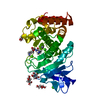
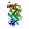
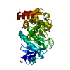
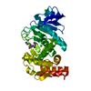
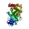
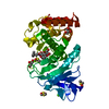
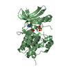
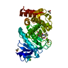
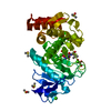
- PDB-1t82: Crystal Structure of the putative thioesterase from Shewanella on... -
+ Open data

- Basic information
| Entry | Database: PDB / ID: 1t82 | ||||||
|---|---|---|---|---|---|---|---|
| Title | Crystal Structure of the putative thioesterase from Shewanella oneidensis, Northeast Structural Genomics Target SoR51 | ||||||
 Components Components | hypothetical acetyltransferase | ||||||
 Keywords Keywords | TRANSFERASE / structural genomics / alpha-beta dimeric protein with a fold resembling a hotdog / PSI / Protein Structure Initiative / Northeast Structural Genomics Consortium / NESG | ||||||
| Function / homology | Thioesterase, putative / Putative thioesterase (yiiD_Cterm) / Hotdog Thioesterase / Thiol Ester Dehydrase; Chain A / acyltransferase activity, transferring groups other than amino-acyl groups / HotDog domain superfamily / Roll / Alpha Beta / Thioesterase domain protein YiiD Function and homology information Function and homology information | ||||||
| Biological species |  Shewanella oneidensis (bacteria) Shewanella oneidensis (bacteria) | ||||||
| Method |  X-RAY DIFFRACTION / SYNCHROTRON / SAD / Resolution: 1.7 Å X-RAY DIFFRACTION / SYNCHROTRON / SAD / Resolution: 1.7 Å | ||||||
 Authors Authors | Forouhar, F. / Lee, I. / Vorobiev, S.M. / Xiao, R. / Acton, T.B. / Montelione, G.T. / Hunt, J.F. / Tong, L. / Northeast Structural Genomics Consortium (NESG) | ||||||
 Citation Citation | Journal: TO BE PUBLISHED Title: Crystal Structure of the putative thioesterase from Shewanella oneidensis, Northeast Structural Genomics Target SoR51 Authors: Forouhar, F. / Lee, I. / Vorobiev, S.M. / Xiao, R. / Acton, T.B. / Montelione, G.T. / Hunt, J.F. / Tong, L. | ||||||
| History |
|
- Structure visualization
| Structure viewer | Molecule: MolmilJmol/JSmol |
|---|
- Downloads & links
-Download
| PDBx/mmCIF format | 1t82.cif.gz | 131.1 KB | Display | PDBx/mmCIF format |
|---|---|---|---|---|
| PDB format | pdb1t82.ent.gz | 102.7 KB | Display | PDB format |
| PDBx/mmJSON format | 1t82.json.gz | Tree view | PDBx/mmJSON format | |
| Others |  Other downloads Other downloads |
-Validation report
| Arichive directory | https://data.pdbj.org/pub/pdb/validation_reports/t8/1t82ftp://data.pdbj.org/pub/pdb/validation_reports/t8/1t82 | HTTPS FTP |
|---|
-Related structure data
| Similar structure data | |
|---|---|
| Other databases |

-Links
 PDBj
PDBj
- Assembly
| Deposited unit | 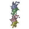
| ||||||||
|---|---|---|---|---|---|---|---|---|---|
| 1 | 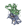
| ||||||||
| 2 | 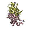
| ||||||||
| Unit cell |
|
-Components
| #1: Protein | Mass: 17851.510 Da / Num. of mol.: 4 Source method: isolated from a genetically manipulated source Source: (gene. exp.) Shewanella oneidensis (bacteria) / Strain: MR-1 / Plasmid: BL21(DE3)+Magic / Species (production host): Escherichia coli / Production host: #2: Water | ChemComp-HOH / |  Mass: 18.015 Da / Num. of mol.: 434 / Source method: isolated from a natural source / Formula: H2O Mass: 18.015 Da / Num. of mol.: 434 / Source method: isolated from a natural source / Formula: H2OHas protein modification | Y | |
|---|
-Experimental details
-Experiment
| Experiment | Method: X-RAY DIFFRACTION / Number of used crystals: 1 |
|---|
- Sample preparation
| Crystal | Density Matthews: 2.1 Å3/Da / Density % sol: 40 % |
|---|---|
| Crystal grow | Temperature: 277 K / Method: vapor diffusion, hanging drop / pH: 7.5 Details: Protein solution: 10mM Tris, 100mM NaCl, 5mM DTT. Reservoir solution: 20% PEG 3350, 200mM sodium formate, pH 7.5, VAPOR DIFFUSION, HANGING DROP, temperature 277K |
-Data collection
| Diffraction | Mean temperature: 100 K |
|---|---|
| Diffraction source | Source: SYNCHROTRON / Site: NSLS  / Beamline: X4A / Wavelength: 0.9793 Å / Beamline: X4A / Wavelength: 0.9793 Å |
| Detector | Type: ADSC QUANTUM 4 / Detector: CCD / Date: Apr 28, 2004 / Details: mirrors |
| Radiation | Monochromator: Si 111 CHANNEL / Protocol: SINGLE WAVELENGTH / Monochromatic (M) / Laue (L): M / Scattering type: x-ray |
| Radiation wavelength | Wavelength: 0.9793 Å / Relative weight: 1 |
| Reflection | Resolution: 1.7→29.81 Å / Num. all: 112723 / Num. obs: 107847 / % possible obs: 92.9 % / Observed criterion σ(F): 2 / Observed criterion σ(I): 2 / Redundancy: 4.43 % / Biso Wilson estimate: 12.4 Å2 / Rmerge(I) obs: 0.07 / Rsym value: 0.079 / Net I/σ(I): 21 |
| Reflection shell | Resolution: 1.7→1.81 Å / Redundancy: 4 % / Rmerge(I) obs: 0.164 / Mean I/σ(I) obs: 9.73 / Num. unique all: 15130 / Rsym value: 0.171 / % possible all: 86.9 |
- Processing
| Software |
| |||||||||||||||||||||||||
|---|---|---|---|---|---|---|---|---|---|---|---|---|---|---|---|---|---|---|---|---|---|---|---|---|---|---|
| Refinement | Method to determine structure: SAD / Resolution: 1.7→29.81 Å Isotropic thermal model: anisotropic and overall temperature factors Cross valid method: THROUGHOUT / σ(F): 2 / σ(I): 2 / Stereochemistry target values: Engh & Huber / Details: Used weighted full matrix least squares procedure.
| |||||||||||||||||||||||||
| Displacement parameters | Biso mean: 15.3 Å2
| |||||||||||||||||||||||||
| Refine analyze |
| |||||||||||||||||||||||||
| Refinement step | Cycle: LAST / Resolution: 1.7→29.81 Å
| |||||||||||||||||||||||||
| Refine LS restraints |
| |||||||||||||||||||||||||
| LS refinement shell | Resolution: 1.7→1.81 Å / Rfactor Rfree error: 0.005
|
