 Movie
Movie Controller

[English] 日本語
 Yorodumi
- PDB-4hgq: Crystal structure of E56A mutant of 2-keto-3-deoxy-D-glycero-D-ga... -
+ Open data

- Basic information
| Entry | Database: PDB / ID: 4hgq | ||||||
|---|---|---|---|---|---|---|---|
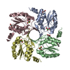
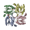
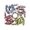
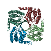
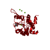
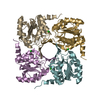
| Title | Crystal structure of E56A mutant of 2-keto-3-deoxy-D-glycero-D-galactonononate-9-phosphate phosphohydrolase from Bacteroides thetaiotaomicron | ||||||
 Components Components | Acylneuraminate cytidylyltransferase | ||||||
 Keywords Keywords | Transferase / Hydrolase / Rossmann Fold / Phosphohydroylase | ||||||
| Function / homology |  Function and homology information Function and homology information3-deoxy-D-glycero-D-galacto-nonulopyranosonate 9-phosphatase / hydrolase activity, acting on ester bonds / metal ion binding Similarity search - Function | ||||||
| Biological species |  Bacteroides thetaiotaomicron (bacteria) Bacteroides thetaiotaomicron (bacteria) | ||||||
| Method |  X-RAY DIFFRACTION / MOLECULAR REPLACEMENT / Resolution: 2.28 Å X-RAY DIFFRACTION / MOLECULAR REPLACEMENT / Resolution: 2.28 Å | ||||||
 Authors Authors | Daughtry, K.D. / Allen, K.N. | ||||||
 Citation Citation | Journal: Biochemistry / Year: 2013 Title: Structural Basis for the Divergence of Substrate Specificity and Biological Function within HAD Phosphatases in Lipopolysaccharide and Sialic Acid Biosynthesis. Authors: Daughtry, K.D. / Huang, H. / Malashkevich, V. / Patskovsky, Y. / Liu, W. / Ramagopal, U. / Sauder, J.M. / Burley, S.K. / Almo, S.C. / Dunaway-Mariano, D. / Allen, K.N. | ||||||
| History |
|
- Structure visualization
| Structure viewer | Molecule: MolmilJmol/JSmol |
|---|
- Downloads & links
-Download
| PDBx/mmCIF format | 4hgq.cif.gz | 272.2 KB | Display | PDBx/mmCIF format |
|---|---|---|---|---|
| PDB format | pdb4hgq.ent.gz | 221.3 KB | Display | PDB format |
| PDBx/mmJSON format | 4hgq.json.gz | Tree view | PDBx/mmJSON format | |
| Others |  Other downloads Other downloads |
-Validation report
| Summary document | 4hgq_validation.pdf.gz | 473.6 KB | Display | wwPDB validaton report |
|---|---|---|---|---|
| Full document | 4hgq_full_validation.pdf.gz | 489.5 KB | Display | |
| Data in XML | 4hgq_validation.xml.gz | 51.9 KB | Display | |
| Data in CIF | 4hgq_validation.cif.gz | 72.6 KB | Display | |
| Arichive directory | https://data.pdbj.org/pub/pdb/validation_reports/hg/4hgqftp://data.pdbj.org/pub/pdb/validation_reports/hg/4hgq | HTTPS FTP |
-Related structure data
| Related structure data |  3mmzC  3mn1C  3n07C  4hgnC  4hgoC  4hgpC  4hgrC  3e8mS S: Starting model for refinement C: citing same article ( |
|---|---|
| Similar structure data |
-Links
 PDBj
PDBj


- Assembly
| Deposited unit | 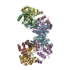
| ||||||||
|---|---|---|---|---|---|---|---|---|---|
| 1 | 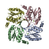
| ||||||||
| 2 | 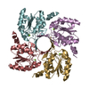
| ||||||||
| Unit cell |
|
-Components
| #1: Protein | Mass: 18304.027 Da / Num. of mol.: 8 / Mutation: E56A Source method: isolated from a genetically manipulated source Source: (gene. exp.) Bacteroides thetaiotaomicron (bacteria)Strain: VPI-5482 / Gene: BT_1713 / Plasmid: pET-3A-626 / Production host: #2: Chemical | ChemComp-MG /   Mass: 24.305 Da / Num. of mol.: 8 / Source method: obtained synthetically / Formula: Mg Mass: 24.305 Da / Num. of mol.: 8 / Source method: obtained synthetically / Formula: Mg#3: Water | ChemComp-HOH / |  Mass: 18.015 Da / Num. of mol.: 562 / Source method: isolated from a natural source / Formula: H2O Mass: 18.015 Da / Num. of mol.: 562 / Source method: isolated from a natural source / Formula: H2O |
|---|
-Experimental details
-Experiment
| Experiment | Method: X-RAY DIFFRACTION / Number of used crystals: 1 |
|---|
- Sample preparation
| Crystal | Density Matthews: 2.31 Å3/Da / Density % sol: 46.71 % |
|---|---|
| Crystal grow | Temperature: 295 K / Method: vapor diffusion, hanging drop / pH: 6.5 Details: 100 mM Bis-Tris, 200 mM magnesium chloride, 23% polyethylene glycol 3350. The crystal was dragged through paratone prior to flash-cooling, pH 6.5, VAPOR DIFFUSION, HANGING DROP, temperature 295K |
-Data collection
| Diffraction | Mean temperature: 100 K |
|---|---|
| Diffraction source | Source: ROTATING ANODE / Type: BRUKER AXS MICROSTAR / Wavelength: 1.54 Å |
| Detector | Type: Bruker Platinum 135 / Detector: CCD / Date: Jul 28, 2009 / Details: Helios multi-layer optics |
| Radiation | Monochromator: rotating-anode / Protocol: SINGLE WAVELENGTH / Monochromatic (M) / Laue (L): M / Scattering type: x-ray |
| Radiation wavelength | Wavelength: 1.54 Å / Relative weight: 1 |
| Reflection | Resolution: 2.28→37.1 Å / Num. all: 62511 / Num. obs: 62511 / % possible obs: 99.9 % / Observed criterion σ(F): 2 / Observed criterion σ(I): 2 / Redundancy: 6.21 % / Rsym value: 0.0878 / Net I/σ(I): 11.6 |
| Reflection shell | Resolution: 2.28→2.37 Å / Redundancy: 4.63 % / Mean I/σ(I) obs: 2.2 / Num. unique all: 3877 / Rsym value: 0.4648 / % possible all: 100 |
- Processing
| Software |
| |||||||||||||||||||||||||||||||||||||||||||||||||||||||||||||||||||||||||||||
|---|---|---|---|---|---|---|---|---|---|---|---|---|---|---|---|---|---|---|---|---|---|---|---|---|---|---|---|---|---|---|---|---|---|---|---|---|---|---|---|---|---|---|---|---|---|---|---|---|---|---|---|---|---|---|---|---|---|---|---|---|---|---|---|---|---|---|---|---|---|---|---|---|---|---|---|---|---|---|
| Refinement | Method to determine structure: MOLECULAR REPLACEMENT Starting model: PDB entry 3e8m Resolution: 2.28→37.1 Å / SU ML: 0.33 / σ(F): 0 / Phase error: 24.57 / Stereochemistry target values: ML
| |||||||||||||||||||||||||||||||||||||||||||||||||||||||||||||||||||||||||||||
| Solvent computation | Shrinkage radii: 0.83 Å / VDW probe radii: 1.1 Å / Solvent model: FLAT BULK SOLVENT MODEL / Bsol: 20.641 Å2 / ksol: 0.316 e/Å3 | |||||||||||||||||||||||||||||||||||||||||||||||||||||||||||||||||||||||||||||
| Displacement parameters | Biso mean: 25.75 Å2
| |||||||||||||||||||||||||||||||||||||||||||||||||||||||||||||||||||||||||||||
| Refine analyze | Luzzati coordinate error obs: 0.33 Å | |||||||||||||||||||||||||||||||||||||||||||||||||||||||||||||||||||||||||||||
| Refinement step | Cycle: LAST / Resolution: 2.28→37.1 Å
| |||||||||||||||||||||||||||||||||||||||||||||||||||||||||||||||||||||||||||||
| Refine LS restraints |
| |||||||||||||||||||||||||||||||||||||||||||||||||||||||||||||||||||||||||||||
| LS refinement shell |
|
