 Movie
Movie Controller

[English] 日本語
 Yorodumi
- PDB-3nrj: Crystal structure of probable yrbi family phosphatase from pseudo... -
+ Open data

- Basic information
| Entry | Database: PDB / ID: 3nrj | ||||||
|---|---|---|---|---|---|---|---|
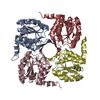
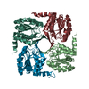
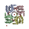
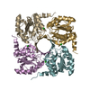
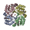
| Title | Crystal structure of probable yrbi family phosphatase from pseudomonas syringae pv.phaseolica 1448a complexed with magnesium | ||||||
 Components Components | PROBABLE YRBI FAMILY PHOSPHATASE | ||||||
 Keywords Keywords | HYDROLASE / STRUCTURAL GENOMICS / PSI / PROTEIN STRUCTURE INITIATIVE / PHOSPHATASE / NEW YORK STRUCTURAL GENOMIX RESEARCH CONSORTIUM / NYSGXRC / NEW YORK SGX RESEARCH CENTER FOR STRUCTURAL GENOMICS | ||||||
| Function / homology |  Function and homology information Function and homology information3-deoxy-manno-octulosonate-8-phosphatase / 3-deoxy-manno-octulosonate-8-phosphatase activity / N-acylneuraminate cytidylyltransferase activity / lipopolysaccharide biosynthetic process / metal ion binding Similarity search - Function | ||||||
| Biological species |  Pseudomonas syringae pv. phaseolicola (bacteria) Pseudomonas syringae pv. phaseolicola (bacteria) | ||||||
| Method |  X-RAY DIFFRACTION / SYNCHROTRON / molecular replacement / Resolution: 1.9 Å X-RAY DIFFRACTION / SYNCHROTRON / molecular replacement / Resolution: 1.9 Å | ||||||
 Authors Authors | Patskovsky, Y. / Ramagopal, U. / Toro, R. / Freeman, J. / Sauder, J.M. / Burley, S.K. / Almo, S.C. / New York SGX Research Center for Structural Genomics (NYSGXRC) | ||||||
 Citation Citation | Journal: To be Published Title: Crystal Structure of Had Family Hydrolase from Pseudomonas Syringae Pv.Phaseolica 1448A Authors: Patskovsky, Y. / Ramagopal, U. / Toro, R. / Freeman, J. / Sauder, J.M. / Burley, S.K. / Almo, S.C. | ||||||
| History |
|
- Structure visualization
| Structure viewer | Molecule: MolmilJmol/JSmol |
|---|
- Downloads & links
-Download
| PDBx/mmCIF format | 3nrj.cif.gz | 426.4 KB | Display | PDBx/mmCIF format |
|---|---|---|---|---|
| PDB format | pdb3nrj.ent.gz | 349.9 KB | Display | PDB format |
| PDBx/mmJSON format | 3nrj.json.gz | Tree view | PDBx/mmJSON format | |
| Others |  Other downloads Other downloads |
-Validation report
| Arichive directory | https://data.pdbj.org/pub/pdb/validation_reports/nr/3nrjftp://data.pdbj.org/pub/pdb/validation_reports/nr/3nrj | HTTPS FTP |
|---|
-Related structure data
| Related structure data |  3mn1S S: Starting model for refinement |
|---|---|
| Similar structure data | |
| Other databases |

-Links
 PDBj
PDBj


- Assembly
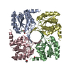
| Deposited unit | 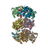
| ||||||||
|---|---|---|---|---|---|---|---|---|---|
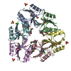
| 1 | 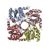
| ||||||||
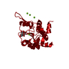
| 2 | 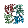
| ||||||||
| 3 | 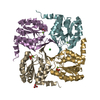
| ||||||||
| Unit cell |
|
-Components
-Protein , 1 types, 12 molecules ABCDEFGHIJKL
| #1: Protein | Mass: 20673.576 Da / Num. of mol.: 12 Source method: isolated from a genetically manipulated source Source: (gene. exp.) Pseudomonas syringae pv. phaseolicola (bacteria)Strain: 1448A / Race 6 / Gene: PSPPH_4147 / Production host: |
|---|
-Non-polymers , 5 types, 833 molecules 


| #2: Chemical | ChemComp-CL /  Mass: 35.453 Da / Num. of mol.: 13 / Source method: obtained synthetically / Formula: Cl Mass: 35.453 Da / Num. of mol.: 13 / Source method: obtained synthetically / Formula: Cl#3: Chemical | ChemComp-MG /  Mass: 24.305 Da / Num. of mol.: 12 / Source method: obtained synthetically / Formula: Mg Mass: 24.305 Da / Num. of mol.: 12 / Source method: obtained synthetically / Formula: Mg#4: Chemical | ChemComp-PO4 /  Mass: 94.971 Da / Num. of mol.: 12 / Source method: obtained synthetically / Formula: PO4 Mass: 94.971 Da / Num. of mol.: 12 / Source method: obtained synthetically / Formula: PO4#5: Chemical | ChemComp-UNL / | Num. of mol.: 1 / Source method: obtained synthetically #6: Water | ChemComp-HOH / | Mass: 18.015 Da / Num. of mol.: 795 / Source method: isolated from a natural source / Formula: H2O |
|---|
-Experimental details
-Experiment
| Experiment | Method: X-RAY DIFFRACTION / Number of used crystals: 1 |
|---|
- Sample preparation
| Crystal | Density Matthews: 2.11 Å3/Da / Density % sol: 41.64 % |
|---|---|
| Crystal grow | Temperature: 294 K / Method: vapor diffusion, sitting drop / pH: 7 Details: 100MM TRIS-HCL, PH 7.0, 30% PEG 3K, 200MM SODIUM CHLORIDE, 10% GLYCEROL, VAPOR DIFFUSION, SITTING DROP, TEMPERATURE 294K |
-Data collection
| Diffraction | Mean temperature: 100 K |
|---|---|
| Diffraction source | Source: SYNCHROTRON / Site: NSLS  / Beamline: X29A / Wavelength: 0.979 / Beamline: X29A / Wavelength: 0.979 |
| Detector | Type: ADSC QUANTUM 315 / Detector: CCD / Date: Apr 29, 2010 / Details: MIRRORS |
| Radiation | Protocol: SINGLE WAVELENGTH / Monochromatic (M) / Laue (L): M / Scattering type: x-ray |
| Radiation wavelength | Wavelength: 0.979 Å / Relative weight: 1 |
| Reflection | Resolution: 1.88→40 Å / Num. obs: 163907 / % possible obs: 95.6 % / Observed criterion σ(I): -5 / Redundancy: 2 % / Biso Wilson estimate: 32.512 Å2 / Rsym value: 0.064 / Net I/σ(I): 6.5 |
| Reflection shell | Resolution: 1.88→1.91 Å / Redundancy: 1.7 % / Rmerge(I) obs: 0.9 / Mean I/σ(I) obs: 0.95 / % possible all: 71.9 |
- Processing
| Software |
| ||||||||||||||||||||||||||||||||||||||||||||||||||||||||||||||||||||||||||||||||||||||||||||||||||||||||||||||||||||||||||||||||||||||||||||||||||||||||||||||||||||||||||
|---|---|---|---|---|---|---|---|---|---|---|---|---|---|---|---|---|---|---|---|---|---|---|---|---|---|---|---|---|---|---|---|---|---|---|---|---|---|---|---|---|---|---|---|---|---|---|---|---|---|---|---|---|---|---|---|---|---|---|---|---|---|---|---|---|---|---|---|---|---|---|---|---|---|---|---|---|---|---|---|---|---|---|---|---|---|---|---|---|---|---|---|---|---|---|---|---|---|---|---|---|---|---|---|---|---|---|---|---|---|---|---|---|---|---|---|---|---|---|---|---|---|---|---|---|---|---|---|---|---|---|---|---|---|---|---|---|---|---|---|---|---|---|---|---|---|---|---|---|---|---|---|---|---|---|---|---|---|---|---|---|---|---|---|---|---|---|---|---|---|---|---|
| Refinement | Method to determine structure: molecular replacement Starting model: 3MN1 Resolution: 1.9→40 Å / Cor.coef. Fo:Fc: 0.974 / Cor.coef. Fo:Fc free: 0.954 / SU B: 5.318 / SU ML: 0.145 / Cross valid method: THROUGHOUT / ESU R: 0.158 / ESU R Free: 0.157 / Stereochemistry target values: MAXIMUM LIKELIHOOD / Details: HYDROGENS HAVE BEEN ADDED IN THE RIDING POSITIONS
| ||||||||||||||||||||||||||||||||||||||||||||||||||||||||||||||||||||||||||||||||||||||||||||||||||||||||||||||||||||||||||||||||||||||||||||||||||||||||||||||||||||||||||
| Solvent computation | Ion probe radii: 0.8 Å / Shrinkage radii: 0.8 Å / VDW probe radii: 1.4 Å / Solvent model: BABINET MODEL WITH MASK | ||||||||||||||||||||||||||||||||||||||||||||||||||||||||||||||||||||||||||||||||||||||||||||||||||||||||||||||||||||||||||||||||||||||||||||||||||||||||||||||||||||||||||
| Displacement parameters | Biso mean: 52.477 Å2
| ||||||||||||||||||||||||||||||||||||||||||||||||||||||||||||||||||||||||||||||||||||||||||||||||||||||||||||||||||||||||||||||||||||||||||||||||||||||||||||||||||||||||||
| Refinement step | Cycle: LAST / Resolution: 1.9→40 Å
| ||||||||||||||||||||||||||||||||||||||||||||||||||||||||||||||||||||||||||||||||||||||||||||||||||||||||||||||||||||||||||||||||||||||||||||||||||||||||||||||||||||||||||
| Refine LS restraints |
| ||||||||||||||||||||||||||||||||||||||||||||||||||||||||||||||||||||||||||||||||||||||||||||||||||||||||||||||||||||||||||||||||||||||||||||||||||||||||||||||||||||||||||
| LS refinement shell | Resolution: 1.9→1.949 Å / Total num. of bins used: 20
|
