 Movie
Movie Controller

[English] 日本語
 Yorodumi
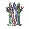
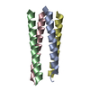
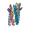
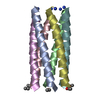
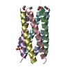
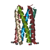
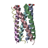
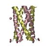
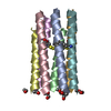
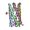
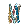
- PDB-4h8m: Crystal structure of a parallel 6-helix coiled coil CC-Hex-H24-A5/7C -
+ Open data

- Basic information
| Entry | Database: PDB / ID: 4h8m | ||||||
|---|---|---|---|---|---|---|---|
| Title | Crystal structure of a parallel 6-helix coiled coil CC-Hex-H24-A5/7C | ||||||
 Components Components | CC-Hex-H24-A5/7C | ||||||
 Keywords Keywords | DE NOVO PROTEIN / CC-Hex / 6-stranded coiled coil / parallel / disulfide bonds / N-terminal acetylation / C-terminal amidation | ||||||
| Biological species | Synthetic construct (others) | ||||||
| Method |  X-RAY DIFFRACTION / SYNCHROTRON / MOLECULAR REPLACEMENT / Resolution: 1.4292 Å X-RAY DIFFRACTION / SYNCHROTRON / MOLECULAR REPLACEMENT / Resolution: 1.4292 Å | ||||||
 Authors Authors | Chi, B. / Zaccai, N.R. / Brady, R.L. / Woolfson, D.N. | ||||||
 Citation Citation | Journal: To be Published Title: TBD Authors: Chi, B. / Zaccai, N.R. / Brady, R.L. / Woolfson, D.N. | ||||||
| History |
|
- Structure visualization
| Structure viewer | Molecule:  MolmilJmol/JSmol MolmilJmol/JSmol |
|---|
- Downloads & links
-Download
| PDBx/mmCIF format | 4h8m.cif.gz | 21.7 KB | Display | PDBx/mmCIF format |
|---|---|---|---|---|
| PDB format | pdb4h8m.ent.gz | 14.9 KB | Display | PDB format |
| PDBx/mmJSON format | 4h8m.json.gz | Tree view | PDBx/mmJSON format | |
| Others |  Other downloads Other downloads |
-Validation report
| Summary document | 4h8m_validation.pdf.gz | 427.2 KB | Display | wwPDB validaton report |
|---|---|---|---|---|
| Full document | 4h8m_full_validation.pdf.gz | 427.7 KB | Display | |
| Data in XML | 4h8m_validation.xml.gz | 4.9 KB | Display | |
| Data in CIF | 4h8m_validation.cif.gz | 6.2 KB | Display | |
| Arichive directory | https://data.pdbj.org/pub/pdb/validation_reports/h8/4h8mftp://data.pdbj.org/pub/pdb/validation_reports/h8/4h8m | HTTPS FTP |
-Related structure data
| Related structure data | 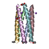 3r46S S: Starting model for refinement |
|---|---|
| Similar structure data |


-Links
 PDBj
PDBj
- Assembly
| Deposited unit | 
| ||||||||||||||||||||||||||||||
|---|---|---|---|---|---|---|---|---|---|---|---|---|---|---|---|---|---|---|---|---|---|---|---|---|---|---|---|---|---|---|---|
| 1 | 
| ||||||||||||||||||||||||||||||
| Unit cell |
| ||||||||||||||||||||||||||||||
| Components on special symmetry positions |
| ||||||||||||||||||||||||||||||
| Details | Disulfide-bridged parallel hexameric coiled coil in redox conditions |
-Components
| #1: Protein/peptide | Mass: 3323.990 Da / Num. of mol.: 2 / Source method: obtained synthetically / Details: standard F-moc solid phase peptide synthesis / Source: (synth.) Synthetic construct (others) #2: Water | ChemComp-HOH / |  Mass: 18.015 Da / Num. of mol.: 56 / Source method: isolated from a natural source / Formula: H2O Mass: 18.015 Da / Num. of mol.: 56 / Source method: isolated from a natural source / Formula: H2OHas protein modification | Y | |
|---|
-Experimental details
-Experiment
| Experiment | Method: X-RAY DIFFRACTION |
|---|
- Sample preparation
| Crystal | Density Matthews: 1.92 Å3/Da / Density % sol: 35.95 % |
|---|---|
| Crystal grow | Temperature: 291 K / Method: vapor diffusion, sitting drop / pH: 7.5 Details: 30 mM sodium fluoride, 30 mM sodium bromide, 30 mM sodium iodide, 50 mM sodium HEPES, 50 mM MOPS (acid) pH 7.5, 20 % PEG-MME 550 and 10 % PEG 20K, VAPOR DIFFUSION, SITTING DROP, temperature 291K |
-Data collection
| Diffraction source | Source: SYNCHROTRON / Site: Diamond  / Beamline: I04 / Wavelength: 0.97 Å / Beamline: I04 / Wavelength: 0.97 Å |
|---|---|
| Detector | Type: ADSC QUANTUM 315 / Detector: CCD / Date: Jul 15, 2011 |
| Radiation | Protocol: SINGLE WAVELENGTH / Monochromatic (M) / Laue (L): M / Scattering type: x-ray |
| Radiation wavelength | Wavelength: 0.97 Å / Relative weight: 1 |
| Reflection | Resolution: 1.429→44.013 Å / Num. all: 9188 / Num. obs: 9078 |
- Processing
| Software | Name: PHENIX / Version: (phenix.refine: 1.7_650) / Classification: refinement | ||||||||||||||||||||||||||||
|---|---|---|---|---|---|---|---|---|---|---|---|---|---|---|---|---|---|---|---|---|---|---|---|---|---|---|---|---|---|
| Refinement | Method to determine structure: MOLECULAR REPLACEMENT Starting model: PDB ENTRY 3R46 Resolution: 1.4292→44.013 Å / SU ML: 0.17 / σ(F): 0.01 / Phase error: 20.6 / Stereochemistry target values: ML
| ||||||||||||||||||||||||||||
| Solvent computation | Shrinkage radii: 0.61 Å / VDW probe radii: 0.9 Å / Solvent model: FLAT BULK SOLVENT MODEL / Bsol: 46.999 Å2 / ksol: 0.374 e/Å3 | ||||||||||||||||||||||||||||
| Displacement parameters |
| ||||||||||||||||||||||||||||
| Refinement step | Cycle: LAST / Resolution: 1.4292→44.013 Å
| ||||||||||||||||||||||||||||
| Refine LS restraints |
| ||||||||||||||||||||||||||||
| LS refinement shell |
|
