 Movie
Movie Controller


+ Open data

- Basic information
| Entry | Database: PDB / ID: 3r3k | ||||||
|---|---|---|---|---|---|---|---|
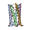
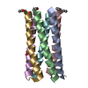
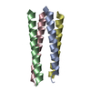
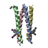
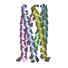
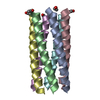
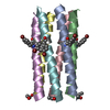
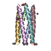
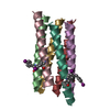
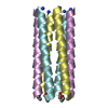
| Title | Crystal structure of a parallel 6-helix coiled coil | ||||||
 Components Components | CChex-Phi22 helix | ||||||
 Keywords Keywords | DE NOVO PROTEIN / parallel hexamer / KIH interactions / hydrophobic channel / synthetic biology | ||||||
| Method |  X-RAY DIFFRACTION / SYNCHROTRON / SAD / Resolution: 2.2009 Å X-RAY DIFFRACTION / SYNCHROTRON / SAD / Resolution: 2.2009 Å | ||||||
 Authors Authors | Zaccai, N.R. / Chi, B.H.C. / Woolfson, D.N. / Brady, R.L. | ||||||
 Citation Citation | Journal: Nat.Chem.Biol. / Year: 2011 Title: A de novo peptide hexamer with a mutable channel. Authors: Zaccai, N.R. / Chi, B. / Thomson, A.R. / Boyle, A.L. / Bartlett, G.J. / Bruning, M. / Linden, N. / Sessions, R.B. / Booth, P.J. / Brady, R.L. / Woolfson, D.N. | ||||||
| History |
|
- Structure visualization
| Structure viewer | Molecule:  MolmilJmol/JSmol MolmilJmol/JSmol |
|---|
- Downloads & links
-Download
| PDBx/mmCIF format | 3r3k.cif.gz | 30 KB | Display | PDBx/mmCIF format |
|---|---|---|---|---|
| PDB format | pdb3r3k.ent.gz | 21.9 KB | Display | PDB format |
| PDBx/mmJSON format | 3r3k.json.gz | Tree view | PDBx/mmJSON format | |
| Others |  Other downloads Other downloads |
-Validation report
| Arichive directory | https://data.pdbj.org/pub/pdb/validation_reports/r3/3r3kftp://data.pdbj.org/pub/pdb/validation_reports/r3/3r3k | HTTPS FTP |
|---|
-Related structure data


-Links
 PDBj
PDBj
- Assembly
| Deposited unit | 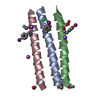
| |||||||||||||||||||||||||||
|---|---|---|---|---|---|---|---|---|---|---|---|---|---|---|---|---|---|---|---|---|---|---|---|---|---|---|---|---|
| 1 | 
| |||||||||||||||||||||||||||
| 2 | 
| |||||||||||||||||||||||||||
| Unit cell |
| |||||||||||||||||||||||||||
| Components on special symmetry positions |
|
-Components
| #1: Protein/peptide | Mass: 3477.891 Da / Num. of mol.: 3 / Fragment: helix from coiled coil domain / Source method: obtained synthetically Details: Peptide synthesis carried out according to standard Fmoc SPPS protocols #2: Chemical |   Mass: 35.453 Da / Num. of mol.: 2 / Source method: obtained synthetically / Formula: Cl Mass: 35.453 Da / Num. of mol.: 2 / Source method: obtained synthetically / Formula: Cl#3: Chemical | ChemComp-NA /   Mass: 22.990 Da / Num. of mol.: 4 / Source method: obtained synthetically / Formula: Na Mass: 22.990 Da / Num. of mol.: 4 / Source method: obtained synthetically / Formula: Na#4: Chemical |   Mass: 62.068 Da / Num. of mol.: 2 / Source method: obtained synthetically / Formula: C2H6O2 Mass: 62.068 Da / Num. of mol.: 2 / Source method: obtained synthetically / Formula: C2H6O2#5: Water | ChemComp-HOH / |  Mass: 18.015 Da / Num. of mol.: 71 / Source method: isolated from a natural source / Formula: H2O Mass: 18.015 Da / Num. of mol.: 71 / Source method: isolated from a natural source / Formula: H2OHas protein modification | Y | |
|---|
-Experimental details
-Experiment
| Experiment | Method: X-RAY DIFFRACTION / Number of used crystals: 1 |
|---|
- Sample preparation
| Crystal | Density Matthews: 2.67 Å3/Da / Density % sol: 53.94 % |
|---|---|
| Crystal grow | Temperature: 291 K / Method: vapor diffusion, sitting drop / pH: 7.5 Details: 20 mM sodium L-glutamate, 20 mM alanine (racemic), 20 mM glycine, 20 mM lysine hydrochloride (racemic), 20 mM serine (racemic), 50 mM sodium HEPES, 50 mM MOPS (acid) pH 7.5, 20 % ethylene ...Details: 20 mM sodium L-glutamate, 20 mM alanine (racemic), 20 mM glycine, 20 mM lysine hydrochloride (racemic), 20 mM serine (racemic), 50 mM sodium HEPES, 50 mM MOPS (acid) pH 7.5, 20 % ethylene glycol,10 % PEG 8K, VAPOR DIFFUSION, SITTING DROP, temperature 291K |
-Data collection
| Diffraction | Mean temperature: 200 K |
|---|---|
| Diffraction source | Source: SYNCHROTRON / Site: Diamond  / Beamline: I04 / Wavelength: 1.7 Å / Beamline: I04 / Wavelength: 1.7 Å |
| Detector | Type: ADSC QUANTUM 315 / Detector: CCD / Date: May 17, 2010 |
| Radiation | Protocol: SINGLE WAVELENGTH / Monochromatic (M) / Laue (L): M / Scattering type: x-ray |
| Radiation wavelength | Wavelength: 1.7 Å / Relative weight: 1 |
| Reflection | Resolution: 2.2→50.14 Å / Num. obs: 5716 / % possible obs: 95.6 % / Redundancy: 5.7 % / Rmerge(I) obs: 0.109 / Net I/σ(I): 9.3 |
| Reflection shell | Resolution: 2.2→2.32 Å / Redundancy: 2.8 % / Rmerge(I) obs: 0.339 / Mean I/σ(I) obs: 2.2 / % possible all: 72.1 |
- Processing
| Software |
| ||||||||||||||||||||||||||||
|---|---|---|---|---|---|---|---|---|---|---|---|---|---|---|---|---|---|---|---|---|---|---|---|---|---|---|---|---|---|
| Refinement | Method to determine structure: SAD / Resolution: 2.2009→23.21 Å / SU ML: 0.25 / σ(F): 0 / Phase error: 24.86 / Stereochemistry target values: ML Details: F(+) and F(-) treated separately during refinement.
| ||||||||||||||||||||||||||||
| Solvent computation | Shrinkage radii: 0.9 Å / VDW probe radii: 1.11 Å / Solvent model: FLAT BULK SOLVENT MODEL / Bsol: 61.818 Å2 / ksol: 0.406 e/Å3 | ||||||||||||||||||||||||||||
| Displacement parameters |
| ||||||||||||||||||||||||||||
| Refinement step | Cycle: LAST / Resolution: 2.2009→23.21 Å
| ||||||||||||||||||||||||||||
| Refine LS restraints |
| ||||||||||||||||||||||||||||
| LS refinement shell |
|
