 Movie
Movie Controller

[English] 日本語
 Yorodumi
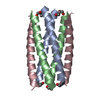
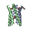
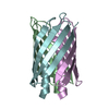
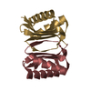
- PDB-6mqu: PL5, synthetic transmembrane domain variant of human phospholamban -
+ Open data

- Basic information
| Entry | Database: PDB / ID: 6mqu | ||||||
|---|---|---|---|---|---|---|---|
| Title | PL5, synthetic transmembrane domain variant of human phospholamban | ||||||
 Components Components | PL5, designed TM pentamer | ||||||
 Keywords Keywords | DE NOVO PROTEIN / designed / engineered / transmembrane / pentamer / phospholamban | ||||||
| Biological species | synthetic construct (others) | ||||||
| Method |  X-RAY DIFFRACTION / SYNCHROTRON / MOLECULAR REPLACEMENT / Resolution: 3.17 Å X-RAY DIFFRACTION / SYNCHROTRON / MOLECULAR REPLACEMENT / Resolution: 3.17 Å | ||||||
 Authors Authors | Mravic, M. / Thomaston, J.L. / DeGrado, W.F. | ||||||
| Funding support |  United States, 1items United States, 1items
| ||||||
 Citation Citation | Journal: Science / Year: 2019 Title: Packing of apolar side chains enables accurate design of highly stable membrane proteins. Authors: Mravic, M. / Thomaston, J.L. / Tucker, M. / Solomon, P.E. / Liu, L. / DeGrado, W.F. | ||||||
| History |
|
- Structure visualization
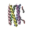
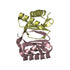
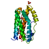
| Structure viewer | Molecule:  MolmilJmol/JSmol MolmilJmol/JSmol |
|---|
- Downloads & links
-Download
| PDBx/mmCIF format | 6mqu.cif.gz | 65.4 KB | Display | PDBx/mmCIF format |
|---|---|---|---|---|
| PDB format | pdb6mqu.ent.gz | 49.4 KB | Display | PDB format |
| PDBx/mmJSON format | 6mqu.json.gz | Tree view | PDBx/mmJSON format | |
| Others |  Other downloads Other downloads |
-Validation report
| Arichive directory | https://data.pdbj.org/pub/pdb/validation_reports/mq/6mquftp://data.pdbj.org/pub/pdb/validation_reports/mq/6mqu | HTTPS FTP |
|---|
-Related structure data
-Links
 PDBj
PDBj

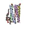
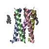
- Assembly
| Deposited unit | 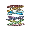
| ||||||||
|---|---|---|---|---|---|---|---|---|---|
| 1 | 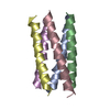
| ||||||||
| 2 | 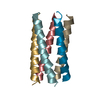
| ||||||||
| Unit cell |
|
-Components
| #1: Protein/peptide | Mass: 3806.718 Da / Num. of mol.: 10 / Source method: obtained synthetically / Source: (synth.) synthetic construct (others) #2: Water | ChemComp-HOH / |  Mass: 18.015 Da / Num. of mol.: 6 / Source method: isolated from a natural source / Formula: H2O Mass: 18.015 Da / Num. of mol.: 6 / Source method: isolated from a natural source / Formula: H2OHas protein modification | N | |
|---|
-Experimental details
-Experiment
| Experiment | Method: X-RAY DIFFRACTION / Number of used crystals: 1 |
|---|
- Sample preparation
| Crystal | Density Matthews: 2.63 Å3/Da / Density % sol: 53.27 % |
|---|---|
| Crystal grow | Temperature: 298 K / Method: vapor diffusion, hanging drop Details: 12 MM C8E4, 20MM MGCL HEXAHYDRATE, 25MM NACL, 50 MM HEPES, 16% PEG400, VAPOR DIFFUSION, TEMPERATURE 298K |
-Data collection
| Diffraction | Mean temperature: 100 K / Serial crystal experiment: N |
|---|---|
| Diffraction source | Source: SYNCHROTRON / Site: ALS / Beamline: 8.3.1 / Wavelength: 1.1158 Å |
| Detector | Type: DECTRIS PILATUS3 S 6M / Detector: PIXEL / Date: May 21, 2017 |
| Radiation | Protocol: SINGLE WAVELENGTH / Monochromatic (M) / Laue (L): M / Scattering type: x-ray |
| Radiation wavelength | Wavelength: 1.1158 Å / Relative weight: 1 |
| Reflection | Resolution: 3.17→87.84 Å / Num. obs: 6819 / % possible obs: 99 % / Redundancy: 3.9 % / Rmerge(I) obs: 0.171 / Net I/σ(I): 6.2 |
| Reflection shell | Resolution: 3.17→3.39 Å / Redundancy: 3.9 % / Rmerge(I) obs: 0.54 / Mean I/σ(I) obs: 2.7 / Num. unique obs: 1212 / CC1/2: 0.802 / Rpim(I) all: 0.314 / % possible all: 98.2 |
- Processing
| Software |
| ||||||||||||||||||||||||||||||||||||||||||
|---|---|---|---|---|---|---|---|---|---|---|---|---|---|---|---|---|---|---|---|---|---|---|---|---|---|---|---|---|---|---|---|---|---|---|---|---|---|---|---|---|---|---|---|
| Refinement | Method to determine structure: MOLECULAR REPLACEMENT / Resolution: 3.17→87.84 Å / SU ML: 0.32 / Cross valid method: FREE R-VALUE / σ(F): 1.34 / Phase error: 25.52
| ||||||||||||||||||||||||||||||||||||||||||
| Solvent computation | Shrinkage radii: 0.9 Å / VDW probe radii: 1.11 Å | ||||||||||||||||||||||||||||||||||||||||||
| Displacement parameters | Biso mean: 46.4 Å2 | ||||||||||||||||||||||||||||||||||||||||||
| Refinement step | Cycle: LAST / Resolution: 3.17→87.84 Å
| ||||||||||||||||||||||||||||||||||||||||||
| Refine LS restraints |
| ||||||||||||||||||||||||||||||||||||||||||
| LS refinement shell |
|
