 Movie
Movie Controller


+ Open data

- Basic information
| Entry | Database: PDB / ID: 5uhs | ||||||
|---|---|---|---|---|---|---|---|
| Title | Structure of a SemiSWEET D57A mutant | ||||||
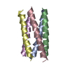
 Components Components | Sugar transporter SemiSWEET | ||||||
 Keywords Keywords | TRANSPORT PROTEIN / membrane / transporter / SemiSWEET | ||||||
| Function / homology |  Function and homology information Function and homology informationD-glucose transmembrane transporter activity / D-glucose transmembrane transport / protein homodimerization activity / membrane / plasma membrane Similarity search - Function | ||||||
| Biological species |  Leptospira biflexa serovar Patoc (bacteria) Leptospira biflexa serovar Patoc (bacteria) | ||||||
| Method |  X-RAY DIFFRACTION / SYNCHROTRON / MOLECULAR REPLACEMENT / Resolution: 2.8 Å X-RAY DIFFRACTION / SYNCHROTRON / MOLECULAR REPLACEMENT / Resolution: 2.8 Å | ||||||
 Authors Authors | Fastman, N.M. / Feng, L. | ||||||
 Citation Citation | Journal: Cell / Year: 2017 Title: Mechanism of Substrate Translocation in an Alternating Access Transporter. Authors: Latorraca, N.R. / Fastman, N.M. / Venkatakrishnan, A.J. / Frommer, W.B. / Dror, R.O. / Feng, L. | ||||||
| History |
|
- Structure visualization
| Structure viewer | Molecule: MolmilJmol/JSmol |
|---|
- Downloads & links
-Download
| PDBx/mmCIF format | 5uhs.cif.gz | 46.8 KB | Display | PDBx/mmCIF format |
|---|---|---|---|---|
| PDB format | pdb5uhs.ent.gz | 31.5 KB | Display | PDB format |
| PDBx/mmJSON format | 5uhs.json.gz | Tree view | PDBx/mmJSON format | |
| Others |  Other downloads Other downloads |
-Validation report
| Arichive directory | https://data.pdbj.org/pub/pdb/validation_reports/uh/5uhsftp://data.pdbj.org/pub/pdb/validation_reports/uh/5uhs | HTTPS FTP |
|---|
-Related structure data
| Related structure data |  5uhqC  4qncS C: citing same article ( S: Starting model for refinement |
|---|---|
| Similar structure data |


-Links
 PDBj
PDBj


- Assembly
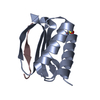
| Deposited unit | 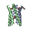
| ||||||||
|---|---|---|---|---|---|---|---|---|---|
| 1 |
| ||||||||
| Unit cell |
|
-Components
| #1: Protein | Mass: 10680.742 Da / Num. of mol.: 2 Source method: isolated from a genetically manipulated source Source: (gene. exp.) Leptospira biflexa serovar Patoc (strain Patoc 1 / ATCC 23582 / Paris) (bacteria)Strain: Patoc 1 / ATCC 23582 / Paris / Gene: LEPBI_I1613 / Production host: #2: Chemical | 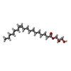  Mass: 356.540 Da / Num. of mol.: 2 / Source method: obtained synthetically / Formula: C21H40O4 Mass: 356.540 Da / Num. of mol.: 2 / Source method: obtained synthetically / Formula: C21H40O4#3: Sugar | ChemComp-BGC / |   Type: D-saccharide, beta linking / Mass: 180.156 Da / Num. of mol.: 1 Type: D-saccharide, beta linking / Mass: 180.156 Da / Num. of mol.: 1Source method: isolated from a genetically manipulated source Formula: C6H12O6 |
|---|
-Experimental details
-Experiment
| Experiment | Method: X-RAY DIFFRACTION / Number of used crystals: 1 |
|---|
- Sample preparation
| Crystal | Density % sol: 34.26 % |
|---|---|
| Crystal grow | Temperature: 293 K / Method: lipidic cubic phase Details: 11.7 MAG 50 mM tris pH 8 100 mM LiSO4 35% PEG 400 40 mM glucose |
-Data collection
| Diffraction | Mean temperature: 100 K |
|---|---|
| Diffraction source | Source: SYNCHROTRON / Site: APS  / Beamline: 23-ID-D / Wavelength: 1.03327 Å / Beamline: 23-ID-D / Wavelength: 1.03327 Å |
| Detector | Type: DECTRIS PILATUS3 6M / Detector: PIXEL / Date: Nov 19, 2014 |
| Radiation | Protocol: SINGLE WAVELENGTH / Monochromatic (M) / Laue (L): M / Scattering type: x-ray |
| Radiation wavelength | Wavelength: 1.03327 Å / Relative weight: 1 |
| Reflection | Resolution: 2.8→50 Å / Num. obs: 4006 / % possible obs: 96.92 % / Redundancy: 9.3 % / Net I/σ(I): 13.73 |
- Processing
| Software |
| ||||||||||||||||||||||||||||
|---|---|---|---|---|---|---|---|---|---|---|---|---|---|---|---|---|---|---|---|---|---|---|---|---|---|---|---|---|---|
| Refinement | Method to determine structure: MOLECULAR REPLACEMENT Starting model: 4qnc Resolution: 2.8→29 Å / SU ML: 0.29 / Cross valid method: FREE R-VALUE / σ(F): 1.34 / Phase error: 30.11
| ||||||||||||||||||||||||||||
| Solvent computation | Shrinkage radii: 0.9 Å / VDW probe radii: 1.11 Å | ||||||||||||||||||||||||||||
| Refinement step | Cycle: LAST / Resolution: 2.8→29 Å
| ||||||||||||||||||||||||||||
| Refine LS restraints |
| ||||||||||||||||||||||||||||
| LS refinement shell |
|
