 Movie
Movie Controller

[English] 日本語
 Yorodumi
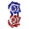
- PDB-4h8k: Crystal structure of LC11-RNase H1 in complex with RNA/DNA hybrid -
+ Open data

- Basic information
| Entry | Database: PDB / ID: 4h8k | ||||||
|---|---|---|---|---|---|---|---|
| Title | Crystal structure of LC11-RNase H1 in complex with RNA/DNA hybrid | ||||||
 Components Components |
| ||||||
 Keywords Keywords | Hydrolase/DNA/RNA / RNase H / Ribonuclease H RNA DNA Hybrid / Hydrolase / Ribonuclease H / Hydrolase-DNA-RNA complex | ||||||
| Function / homology |  Function and homology information Function and homology informationribonuclease H / RNA-DNA hybrid ribonuclease activity / nucleic acid binding Similarity search - Function | ||||||
| Biological species | uncultured organism (environmental samples) | ||||||
| Method |  X-RAY DIFFRACTION / SYNCHROTRON / MOLECULAR REPLACEMENT / Resolution: 2.3 Å X-RAY DIFFRACTION / SYNCHROTRON / MOLECULAR REPLACEMENT / Resolution: 2.3 Å | ||||||
 Authors Authors | Nguyen, T.N. / You, D.J. / Matsumoto, H. / Kanaya, E. / Kanaya, S. | ||||||
 Citation Citation | Journal: J.Struct.Biol. / Year: 2013 Title: Crystal structure of metagenome-derived LC11-RNase H1 in complex with RNA/DNA hybrid Authors: Nguyen, T.N. / You, D.J. / Matsumoto, H. / Kanaya, E. / Koga, Y. / Kanaya, S. | ||||||
| History |
|
- Structure visualization
| Structure viewer | Molecule: MolmilJmol/JSmol |
|---|
- Downloads & links
-Download
| PDBx/mmCIF format | 4h8k.cif.gz | 86.3 KB | Display | PDBx/mmCIF format |
|---|---|---|---|---|
| PDB format | pdb4h8k.ent.gz | 61.2 KB | Display | PDB format |
| PDBx/mmJSON format | 4h8k.json.gz | Tree view | PDBx/mmJSON format | |
| Others |  Other downloads Other downloads |
-Validation report
| Summary document | 4h8k_validation.pdf.gz | 460.2 KB | Display | wwPDB validaton report |
|---|---|---|---|---|
| Full document | 4h8k_full_validation.pdf.gz | 474.6 KB | Display | |
| Data in XML | 4h8k_validation.xml.gz | 15.6 KB | Display | |
| Data in CIF | 4h8k_validation.cif.gz | 21.5 KB | Display | |
| Arichive directory | https://data.pdbj.org/pub/pdb/validation_reports/h8/4h8kftp://data.pdbj.org/pub/pdb/validation_reports/h8/4h8k | HTTPS FTP |
-Related structure data
| Related structure data |  3u3gS S: Starting model for refinement |
|---|---|
| Similar structure data |


-Links
 PDBj
PDBj


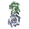
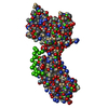
- Assembly
| Deposited unit | 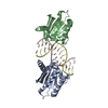
| ||||||||
|---|---|---|---|---|---|---|---|---|---|
| 1 |
| ||||||||
| Unit cell |
|
-Components
| #1: Protein | Mass: 15630.956 Da / Num. of mol.: 2 / Mutation: D77N Source method: isolated from a genetically manipulated source Source: (gene. exp.) uncultured organism (environmental samples) Plasmid: pET25b / Production host:  #2: RNA chain | | Mass: 4382.659 Da / Num. of mol.: 1 / Source method: obtained synthetically / Details: This sequence occurs naturally in bacteria #3: DNA chain | | Mass: 4360.840 Da / Num. of mol.: 1 / Source method: obtained synthetically / Details: This sequence occurs naturally in bacteria #4: Water | ChemComp-HOH / |  Mass: 18.015 Da / Num. of mol.: 92 / Source method: isolated from a natural source / Formula: H2O Mass: 18.015 Da / Num. of mol.: 92 / Source method: isolated from a natural source / Formula: H2O |
|---|
-Experimental details
-Experiment
| Experiment | Method: X-RAY DIFFRACTION / Number of used crystals: 1 |
|---|
- Sample preparation
| Crystal | Density Matthews: 2.66 Å3/Da / Density % sol: 53.81 % |
|---|---|
| Crystal grow | Temperature: 293 K / Method: vapor diffusion, sitting drop / pH: 8.5 Details: 0.2M ammonium acetate, 0.1M Tris, 25% PEG3350, pH 8.5, VAPOR DIFFUSION, SITTING DROP, temperature 293K |
-Data collection
| Diffraction | Mean temperature: 100 K | |||||||||||||||||||||||||||||||||||||||||||||||||
|---|---|---|---|---|---|---|---|---|---|---|---|---|---|---|---|---|---|---|---|---|---|---|---|---|---|---|---|---|---|---|---|---|---|---|---|---|---|---|---|---|---|---|---|---|---|---|---|---|---|---|
| Diffraction source | Source: SYNCHROTRON / Site: SPring-8  / Beamline: BL44XU / Wavelength: 0.9 Å / Beamline: BL44XU / Wavelength: 0.9 Å | |||||||||||||||||||||||||||||||||||||||||||||||||
| Detector | Type: BRUKER SMART 6500 / Detector: CCD / Date: Nov 13, 2011 | |||||||||||||||||||||||||||||||||||||||||||||||||
| Radiation | Monochromator: horizontal focusing mirror / Protocol: SINGLE WAVELENGTH / Monochromatic (M) / Laue (L): M / Scattering type: x-ray | |||||||||||||||||||||||||||||||||||||||||||||||||
| Radiation wavelength | Wavelength: 0.9 Å / Relative weight: 1 | |||||||||||||||||||||||||||||||||||||||||||||||||
| Reflection | Resolution: 2.3→50 Å / Num. obs: 17868 / % possible obs: 95.4 % / Observed criterion σ(F): 0 / Observed criterion σ(I): -3 / Redundancy: 7.4 % / Rmerge(I) obs: 0.049 / Rsym value: 0.049 / Net I/σ(I): 42.556 | |||||||||||||||||||||||||||||||||||||||||||||||||
| Reflection shell |
|
- Processing
| Software |
| ||||||||||||||||||||||||||||||||||||
|---|---|---|---|---|---|---|---|---|---|---|---|---|---|---|---|---|---|---|---|---|---|---|---|---|---|---|---|---|---|---|---|---|---|---|---|---|---|
| Refinement | Method to determine structure: MOLECULAR REPLACEMENT Starting model: 3U3G Resolution: 2.3→38.28 Å / Cor.coef. Fo:Fc: 0.951 / Cor.coef. Fo:Fc free: 0.908 / SU B: 7.64 / SU ML: 0.187 / Cross valid method: THROUGHOUT / σ(F): 0 / ESU R: 0.349 / ESU R Free: 0.27 / Stereochemistry target values: MAXIMUM LIKELIHOOD / Details: HYDROGENS HAVE BEEN ADDED IN THE RIDING POSITIONS
| ||||||||||||||||||||||||||||||||||||
| Solvent computation | Ion probe radii: 0.8 Å / Shrinkage radii: 0.8 Å / VDW probe radii: 1.4 Å / Solvent model: MASK | ||||||||||||||||||||||||||||||||||||
| Displacement parameters | Biso mean: 52.048 Å2
| ||||||||||||||||||||||||||||||||||||
| Refinement step | Cycle: LAST / Resolution: 2.3→38.28 Å
| ||||||||||||||||||||||||||||||||||||
| Refine LS restraints |
| ||||||||||||||||||||||||||||||||||||
| LS refinement shell | Resolution: 2.299→2.359 Å / Total num. of bins used: 20
|
