 Movie
Movie Controller

[English] 日本語
 Yorodumi
- PDB-5h36: Crystal structures of the TRIC trimeric intracellular cation chan... -
+ Open data

- Basic information
| Entry | Database: PDB / ID: 5h36 | ||||||
|---|---|---|---|---|---|---|---|
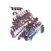
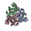
| Title | Crystal structures of the TRIC trimeric intracellular cation channel orthologue from Rhodobacter sphaeroides | ||||||
 Components Components | Uncharacterized protein TRIC | ||||||
 Keywords Keywords | MEMBRANE PROTEIN / ion channels | ||||||
| Function / homology | Glycine transporter / Glycine transporter / plasma membrane / 1,2-DIMYRISTOYL-SN-GLYCERO-3-PHOSPHOCHOLINE / Glycine transporter domain-containing protein Function and homology information Function and homology information | ||||||
| Biological species |  Rhodobacter sphaeroides (bacteria) Rhodobacter sphaeroides (bacteria) | ||||||
| Method |  X-RAY DIFFRACTION / SYNCHROTRON / MAD / Resolution: 3.409 Å X-RAY DIFFRACTION / SYNCHROTRON / MAD / Resolution: 3.409 Å | ||||||
 Authors Authors | Kasuya, G. / Hiraizumi, M. / Hattori, M. / Nureki, O. | ||||||
 Citation Citation | Journal: Cell Res. / Year: 2016 Title: Crystal structures of the TRIC trimeric intracellular cation channel orthologues Authors: Kasuya, G. / Hiraizumi, M. / Maturana, A.D. / Kumazaki, K. / Fujiwara, Y. / Liu, K. / Nakada-Nakura, Y. / Iwata, S. / Tsukada, K. / Komori, T. / Uemura, S. / Goto, Y. / Nakane, T. / ...Authors: Kasuya, G. / Hiraizumi, M. / Maturana, A.D. / Kumazaki, K. / Fujiwara, Y. / Liu, K. / Nakada-Nakura, Y. / Iwata, S. / Tsukada, K. / Komori, T. / Uemura, S. / Goto, Y. / Nakane, T. / Takemoto, M. / Kato, H.E. / Yamashita, K. / Wada, M. / Ito, K. / Ishitani, R. / Hattori, M. / Nureki, O. | ||||||
| History |
|
- Structure visualization
| Structure viewer | Molecule: MolmilJmol/JSmol |
|---|
- Downloads & links
-Download
| PDBx/mmCIF format | 5h36.cif.gz | 84.7 KB | Display | PDBx/mmCIF format |
|---|---|---|---|---|
| PDB format | pdb5h36.ent.gz | 62.6 KB | Display | PDB format |
| PDBx/mmJSON format | 5h36.json.gz | Tree view | PDBx/mmJSON format | |
| Others |  Other downloads Other downloads |
-Validation report
| Arichive directory | https://data.pdbj.org/pub/pdb/validation_reports/h3/5h36ftp://data.pdbj.org/pub/pdb/validation_reports/h3/5h36 | HTTPS FTP |
|---|
-Related structure data


-Links
 PDBj
PDBj

- Assembly
| Deposited unit | 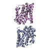
| ||||||||
|---|---|---|---|---|---|---|---|---|---|
| 1 | 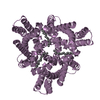
| ||||||||
| 2 | 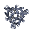
| ||||||||
| Unit cell |
|
-Components
| #1: Protein | Mass: 22783.598 Da / Num. of mol.: 2 / Fragment: UNP residues 1-204 Source method: isolated from a genetically manipulated source Source: (gene. exp.) Rhodobacter sphaeroides (strain ATCC 17023 / 2.4.1 / NCIB 8253 / DSM 158) (bacteria)Strain: ATCC 17023 / 2.4.1 / NCIB 8253 / DSM 158 / Gene: RSP_3856 / Production host: #2: Chemical |   Mass: 678.940 Da / Num. of mol.: 2 / Source method: obtained synthetically / Formula: C36H73NO8P / Comment: DMPC, phospholipid*YM Mass: 678.940 Da / Num. of mol.: 2 / Source method: obtained synthetically / Formula: C36H73NO8P / Comment: DMPC, phospholipid*YM |
|---|
-Experimental details
-Experiment
| Experiment | Method: X-RAY DIFFRACTION / Number of used crystals: 1 |
|---|
- Sample preparation
| Crystal | Density Matthews: 4.23 Å3/Da / Density % sol: 70.95 % |
|---|---|
| Crystal grow | Temperature: 293 K / Method: vapor diffusion Details: 25-32% (v/v) PEG 400, 20-70mM Li2SO4, 20-70mM Na2SO4, 50mM Tris-HCl, pH 8.0 |
-Data collection
| Diffraction | Mean temperature: 100 K |
|---|---|
| Diffraction source | Source: SYNCHROTRON / Site: SLS  / Beamline: X06SA / Wavelength: 1 Å / Beamline: X06SA / Wavelength: 1 Å |
| Detector | Type: PSI PILATUS 6M / Detector: PIXEL / Date: Aug 21, 2014 |
| Radiation | Protocol: SINGLE WAVELENGTH / Monochromatic (M) / Laue (L): M / Scattering type: x-ray |
| Radiation wavelength | Wavelength: 1 Å / Relative weight: 1 |
| Reflection | Resolution: 3.409→50 Å / Num. obs: 9730 / % possible obs: 95.3 % / Redundancy: 5.1 % / Net I/σ(I): 15.2 |
- Processing
| Software |
| ||||||||||||||||||||||||||||||||||||||||||||||||||||||||
|---|---|---|---|---|---|---|---|---|---|---|---|---|---|---|---|---|---|---|---|---|---|---|---|---|---|---|---|---|---|---|---|---|---|---|---|---|---|---|---|---|---|---|---|---|---|---|---|---|---|---|---|---|---|---|---|---|---|
| Refinement | Method to determine structure: MAD / Resolution: 3.409→45.036 Å / Cross valid method: FREE R-VALUE / σ(F): 1.97 / Phase error: 34.53
| ||||||||||||||||||||||||||||||||||||||||||||||||||||||||
| Solvent computation | Shrinkage radii: 0.9 Å / VDW probe radii: 1.11 Å | ||||||||||||||||||||||||||||||||||||||||||||||||||||||||
| Refinement step | Cycle: LAST / Resolution: 3.409→45.036 Å
| ||||||||||||||||||||||||||||||||||||||||||||||||||||||||
| Refine LS restraints |
| ||||||||||||||||||||||||||||||||||||||||||||||||||||||||
| LS refinement shell |
|
