 Movie
Movie Controller

[English] 日本語
 Yorodumi
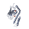
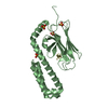
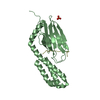
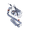
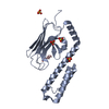
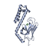
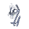
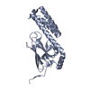
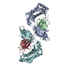
- PDB-4ezu: Crystal structure of the substrate binding domain of E.coli DnaK ... -
+ Open data

- Basic information
| Entry | Database: PDB / ID: 4ezu | ||||||
|---|---|---|---|---|---|---|---|
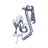
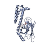
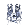
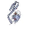
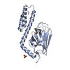
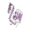
| Title | Crystal structure of the substrate binding domain of E.coli DnaK in complex with PR-bombesin in space group I222 | ||||||
 Components Components |
| ||||||
 Keywords Keywords | CHAPERONE/PEPTIDE BINDING PROTEIN / chaperone / peptide binding / CHAPERONE-PEPTIDE BINDING PROTEIN complex | ||||||
| Function / homology |  Function and homology information Function and homology informationstress response to copper ion / sigma factor antagonist activity / protein unfolding / cellular response to unfolded protein / heat shock protein binding / protein folding chaperone / inclusion body / ATP-dependent protein folding chaperone / neuropeptide signaling pathway / ADP binding ...stress response to copper ion / sigma factor antagonist activity / protein unfolding / cellular response to unfolded protein / heat shock protein binding / protein folding chaperone / inclusion body / ATP-dependent protein folding chaperone / neuropeptide signaling pathway / ADP binding / : / protein refolding / response to heat / protein-folding chaperone binding / protein folding / protein-containing complex assembly / DNA replication / ATP hydrolysis activity / protein-containing complex / extracellular region / zinc ion binding / ATP binding / membrane / plasma membrane / cytosol / cytoplasm Similarity search - Function | ||||||
| Biological species |  Bombina maxima (large-webbed bell toad) | ||||||
| Method |  X-RAY DIFFRACTION / SYNCHROTRON / PDB ID 1DKZ / Resolution: 1.9 Å X-RAY DIFFRACTION / SYNCHROTRON / PDB ID 1DKZ / Resolution: 1.9 Å | ||||||
 Authors Authors | Zahn, M. / Straeter, N. | ||||||
 Citation Citation | Journal: To be Published Title: Structural studies of DnaK in complex with proline rich antimicrobial peptides reveal two different peptide binding modes Authors: Zahn, M. / Straeter, N. | ||||||
| History |
|
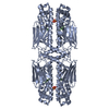
- Structure visualization
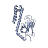
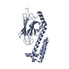
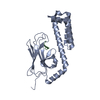
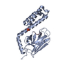
| Structure viewer | Molecule: MolmilJmol/JSmol |
|---|
- Downloads & links
-Download
| PDBx/mmCIF format | 4ezu.cif.gz | 99.4 KB | Display | PDBx/mmCIF format |
|---|---|---|---|---|
| PDB format | pdb4ezu.ent.gz | 78 KB | Display | PDB format |
| PDBx/mmJSON format | 4ezu.json.gz | Tree view | PDBx/mmJSON format | |
| Others |  Other downloads Other downloads |
-Validation report
| Arichive directory | https://data.pdbj.org/pub/pdb/validation_reports/ez/4ezuftp://data.pdbj.org/pub/pdb/validation_reports/ez/4ezu | HTTPS FTP |
|---|
-Related structure data


-Links
 PDBj
PDBj
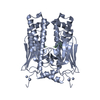
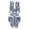
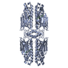
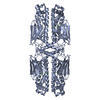
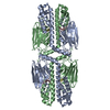
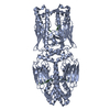
- Assembly
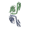
| Deposited unit | 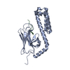
| ||||||||
|---|---|---|---|---|---|---|---|---|---|
| 1 | 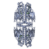
| ||||||||
| 2 | 
| ||||||||
| 3 | 
| ||||||||
| 4 |
| ||||||||
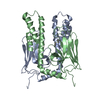
| Unit cell |
|
-Components
| #1: Protein | Mass: 23820.777 Da / Num. of mol.: 1 / Fragment: UNP residues 389-607 Source method: isolated from a genetically manipulated source Source: (gene. exp.) |
|---|---|
| #2: Protein/peptide | Mass: 2040.453 Da / Num. of mol.: 1 / Fragment: UNP residues 46-60 / Source method: obtained synthetically Details: This sequence occurs naturally in Bombina maxima. C-terminus is amidated. Source: (synth.) Bombina maxima (large-webbed bell toad) / References: UniProt: Q8QFP2 |
| #3: Water | ChemComp-HOH /  Mass: 18.015 Da / Num. of mol.: 80 / Source method: isolated from a natural source / Formula: H2O Mass: 18.015 Da / Num. of mol.: 80 / Source method: isolated from a natural source / Formula: H2O |
-Experimental details
-Experiment
| Experiment | Method: X-RAY DIFFRACTION / Number of used crystals: 1 |
|---|
- Sample preparation
| Crystal | Density Matthews: 2.11 Å3/Da / Density % sol: 41.64 % |
|---|---|
| Crystal grow | Temperature: 292 K / Method: vapor diffusion, hanging drop / pH: 8 Details: 3.0 M ammonium sulfate, 0.1 M HEPES pH 8.0, VAPOR DIFFUSION, HANGING DROP, temperature 292K |
-Data collection
| Diffraction | Mean temperature: 100 K |
|---|---|
| Diffraction source | Source: SYNCHROTRON / Site: BESSY  / Beamline: 14.2 / Wavelength: 0.91841 Å / Beamline: 14.2 / Wavelength: 0.91841 Å |
| Detector | Type: MARMOSAIC 225 mm CCD / Detector: CCD / Date: Oct 19, 2010 |
| Radiation | Monochromator: Si - 111 / Protocol: SINGLE WAVELENGTH / Monochromatic (M) / Laue (L): M / Scattering type: x-ray |
| Radiation wavelength | Wavelength: 0.91841 Å / Relative weight: 1 |
| Reflection | Resolution: 1.9→25 Å / Num. obs: 17741 / % possible obs: 99.8 % / Rmerge(I) obs: 0.062 |
| Reflection shell | Resolution: 1.9→2 Å / Redundancy: 5.3 % / Rmerge(I) obs: 0.558 / % possible all: 100 |
- Processing
| Software |
| ||||||||||||||||||||||||||||||||||||||||||||||||||||||||||||||||||||||||||||||||||||||||||||||||||||||||||||||||||||||||||||||||||||||||||||||||||||||||||||||||||||||||||
|---|---|---|---|---|---|---|---|---|---|---|---|---|---|---|---|---|---|---|---|---|---|---|---|---|---|---|---|---|---|---|---|---|---|---|---|---|---|---|---|---|---|---|---|---|---|---|---|---|---|---|---|---|---|---|---|---|---|---|---|---|---|---|---|---|---|---|---|---|---|---|---|---|---|---|---|---|---|---|---|---|---|---|---|---|---|---|---|---|---|---|---|---|---|---|---|---|---|---|---|---|---|---|---|---|---|---|---|---|---|---|---|---|---|---|---|---|---|---|---|---|---|---|---|---|---|---|---|---|---|---|---|---|---|---|---|---|---|---|---|---|---|---|---|---|---|---|---|---|---|---|---|---|---|---|---|---|---|---|---|---|---|---|---|---|---|---|---|---|---|---|---|
| Refinement | Method to determine structure: PDB ID 1DKZ / Resolution: 1.9→25 Å / Cor.coef. Fo:Fc: 0.948 / Cor.coef. Fo:Fc free: 0.916 / SU B: 9.73 / SU ML: 0.145 / Cross valid method: THROUGHOUT / ESU R: 0.187 / ESU R Free: 0.177 / Stereochemistry target values: MAXIMUM LIKELIHOOD / Details: HYDROGENS HAVE BEEN USED IF PRESENT IN THE INPUT
| ||||||||||||||||||||||||||||||||||||||||||||||||||||||||||||||||||||||||||||||||||||||||||||||||||||||||||||||||||||||||||||||||||||||||||||||||||||||||||||||||||||||||||
| Solvent computation | Ion probe radii: 0.8 Å / Shrinkage radii: 0.8 Å / VDW probe radii: 1.2 Å / Solvent model: MASK | ||||||||||||||||||||||||||||||||||||||||||||||||||||||||||||||||||||||||||||||||||||||||||||||||||||||||||||||||||||||||||||||||||||||||||||||||||||||||||||||||||||||||||
| Displacement parameters | Biso mean: 35.847 Å2
| ||||||||||||||||||||||||||||||||||||||||||||||||||||||||||||||||||||||||||||||||||||||||||||||||||||||||||||||||||||||||||||||||||||||||||||||||||||||||||||||||||||||||||
| Refinement step | Cycle: LAST / Resolution: 1.9→25 Å
| ||||||||||||||||||||||||||||||||||||||||||||||||||||||||||||||||||||||||||||||||||||||||||||||||||||||||||||||||||||||||||||||||||||||||||||||||||||||||||||||||||||||||||
| Refine LS restraints |
| ||||||||||||||||||||||||||||||||||||||||||||||||||||||||||||||||||||||||||||||||||||||||||||||||||||||||||||||||||||||||||||||||||||||||||||||||||||||||||||||||||||||||||
| LS refinement shell | Resolution: 1.9→1.949 Å / Total num. of bins used: 20
| ||||||||||||||||||||||||||||||||||||||||||||||||||||||||||||||||||||||||||||||||||||||||||||||||||||||||||||||||||||||||||||||||||||||||||||||||||||||||||||||||||||||||||
| Refinement TLS params. | Method: refined / Refine-ID: X-RAY DIFFRACTION
| ||||||||||||||||||||||||||||||||||||||||||||||||||||||||||||||||||||||||||||||||||||||||||||||||||||||||||||||||||||||||||||||||||||||||||||||||||||||||||||||||||||||||||
| Refinement TLS group |
|
