[English] 日本語
Yorodumi
- PDB-4ezs: Crystal structure of the substrate binding domain of E.coli DnaK ... -

+
Open data


ID or keywords:

Loading...

-
Basic information

Entry
Database: PDB / ID: 4ezs
TitleCrystal structure of the substrate binding domain of E.coli DnaK in complex with metchnikowin (residues 20 to 26)
Components
  • Chaperone protein DnaK
  • Metchnikowin
KeywordsCHAPERONE/PEPTIDE BINDING PROTEIN / chaperone / peptide binding / CHAPERONE-PEPTIDE BINDING PROTEIN complex
Function / homology
Function and homology information


stress response to copper ion / sigma factor antagonist activity / antifungal humoral response / chaperone cofactor-dependent protein refolding / protein unfolding / cellular response to unfolded protein / defense response to fungus / inclusion body / protein folding chaperone / heat shock protein binding ...stress response to copper ion / sigma factor antagonist activity / antifungal humoral response / chaperone cofactor-dependent protein refolding / protein unfolding / cellular response to unfolded protein / defense response to fungus / inclusion body / protein folding chaperone / heat shock protein binding / ADP binding / response to bacterium / ATP-dependent protein folding chaperone / defense response / response to wounding / unfolded protein binding / antibacterial humoral response / response to heat / protein-folding chaperone binding / protein refolding / protein-containing complex assembly / killing of cells of another organism / defense response to Gram-negative bacterium / DNA replication / defense response to Gram-positive bacterium / innate immune response / ATP hydrolysis activity / protein-containing complex / zinc ion binding / extracellular region / ATP binding / membrane / plasma membrane / cytosol / cytoplasm
Similarity search - Function
Metchnikowin / Metchnikowin family / Substrate Binding Domain Of Dnak; Chain:A; Domain 2 - #10 / Substrate Binding Domain Of DNAk; Chain A, domain 1 / Chaperone DnaK / Substrate Binding Domain Of DNAk; Chain A, domain 1 / Heat shock hsp70 proteins family signature 2. / Heat shock hsp70 proteins family signature 1. / Heat shock hsp70 proteins family signature 3. / Heat shock protein 70, conserved site ...Metchnikowin / Metchnikowin family / Substrate Binding Domain Of Dnak; Chain:A; Domain 2 - #10 / Substrate Binding Domain Of DNAk; Chain A, domain 1 / Chaperone DnaK / Substrate Binding Domain Of DNAk; Chain A, domain 1 / Heat shock hsp70 proteins family signature 2. / Heat shock hsp70 proteins family signature 1. / Heat shock hsp70 proteins family signature 3. / Heat shock protein 70, conserved site / Heat shock protein 70kD, peptide-binding domain superfamily / Heat shock protein 70 family / Hsp70 protein / Heat shock protein 70kD, C-terminal domain superfamily / Substrate Binding Domain Of Dnak; Chain:A; Domain 2 / ATPase, nucleotide binding domain / Up-down Bundle / Sandwich / Mainly Beta / Mainly Alpha
Similarity search - Domain/homology
Chaperone protein DnaK / Metchnikowin
Similarity search - Component
Biological speciesEscherichia coli (E. coli)
Drosophila melanogaster (fruit fly)
MethodX-RAY DIFFRACTION / SYNCHROTRON / PDB ENTRY 1DKZ / Resolution: 1.9 Å
AuthorsZahn, M. / Straeter, N.
Citation
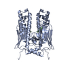
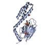
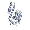
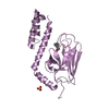
Journal: To be Published
Title: Structural studies of DnaK in complex with proline rich antimicrobial peptides reveal two different peptide binding modes
Authors: Zahn, M. / Straeter, N.
History
DepositionMay 3, 2012Deposition site: RCSB / Processing site: RCSB
Revision 1.0Apr 24, 2013Provider: repository / Type: Initial release
Revision 1.1Nov 15, 2017Group: Refinement description / Category: software / Item: _software.name
Revision 1.2Feb 28, 2024Group: Data collection / Database references / Derived calculations
Category: chem_comp_atom / chem_comp_bond ...chem_comp_atom / chem_comp_bond / database_2 / struct_ref_seq_dif / struct_site
Item: _database_2.pdbx_DOI / _database_2.pdbx_database_accession ..._database_2.pdbx_DOI / _database_2.pdbx_database_accession / _struct_ref_seq_dif.details / _struct_site.pdbx_auth_asym_id / _struct_site.pdbx_auth_comp_id / _struct_site.pdbx_auth_seq_id

-
Structure visualization

Structure viewerMolecule:
MolmilJmol/JSmol

Downloads & links

-
Assembly

Deposited unit
A: Chaperone protein DnaK
B: Metchnikowin
hetero molecules


Theoretical massNumber of molelcules
Total (without water)24,8803
Polymers24,7842
Non-polymers961
Water1,53185
1
A: Chaperone protein DnaK
B: Metchnikowin
hetero molecules

A: Chaperone protein DnaK
B: Metchnikowin
hetero molecules

A: Chaperone protein DnaK
B: Metchnikowin
hetero molecules

A: Chaperone protein DnaK
B: Metchnikowin
hetero molecules


Theoretical massNumber of molelcules
Total (without water)99,52012
Polymers99,1368
Non-polymers3844
Water1448
TypeNameSymmetry operationNumber
identity operation1_555x,y,z1
crystal symmetry operation2_675-x+1,-y+2,z1
crystal symmetry operation3_657-x+1,y,-z+21
crystal symmetry operation4_577x,-y+2,-z+21
Buried area18570 Å2
ΔGint-119 kcal/mol
Surface area36910 Å2
MethodPISA
2
A: Chaperone protein DnaK
B: Metchnikowin
hetero molecules

A: Chaperone protein DnaK
B: Metchnikowin
hetero molecules


Theoretical massNumber of molelcules
Total (without water)49,7606
Polymers49,5684
Non-polymers1922
Water724
TypeNameSymmetry operationNumber
identity operation1_555x,y,z1
crystal symmetry operation3_657-x+1,y,-z+21
Buried area6920 Å2
ΔGint-43 kcal/mol
Surface area20820 Å2
MethodPISA
3
A: Chaperone protein DnaK
B: Metchnikowin
hetero molecules

A: Chaperone protein DnaK
B: Metchnikowin
hetero molecules


Theoretical massNumber of molelcules
Total (without water)49,7606
Polymers49,5684
Non-polymers1922
Water724
TypeNameSymmetry operationNumber
identity operation1_555x,y,z1
crystal symmetry operation4_577x,-y+2,-z+21
Buried area4540 Å2
ΔGint-44 kcal/mol
Surface area23200 Å2
MethodPISA
4
A: Chaperone protein DnaK
B: Metchnikowin
hetero molecules

A: Chaperone protein DnaK
B: Metchnikowin
hetero molecules


Theoretical massNumber of molelcules
Total (without water)49,7606
Polymers49,5684
Non-polymers1922
Water724
TypeNameSymmetry operationNumber
identity operation1_555x,y,z1
crystal symmetry operation3_656-x+1,y,-z+11
Buried area4380 Å2
ΔGint-40 kcal/mol
Surface area23360 Å2
MethodPISA
5


  • Idetical with deposited unit
  • defined by author&software
TypeNameSymmetry operationNumber
identity operation1_555x,y,z1
Buried area1320 Å2
ΔGint-15 kcal/mol
Surface area12550 Å2
MethodPISA
Unit cell
Length a, b, c (Å)94.437, 116.905, 36.994
Angle α, β, γ (deg.)90.00, 90.00, 90.00
Int Tables number23
Space group name H-MI222
Components on special symmetry positions
IDModelComponents
11A-701-

SO4

-
Components

#1: Protein Chaperone protein DnaK / / HSP70 / Heat shock 70 kDa protein / Heat shock protein 70


Mass: 23820.777 Da / Num. of mol.: 1 / Fragment: UNP residues 389-607
Source method: isolated from a genetically manipulated source
Source: (gene. exp.) Escherichia coli (E. coli) / Strain: K12 / Gene: b0014, dnaK, groP, grpF, JW0013, seg / Production host: Escherichia coli (E. coli) / References: UniProt: P0A6Y8
#2: Protein/peptide Metchnikowin /


Mass: 963.111 Da / Num. of mol.: 1 / Fragment: UNP residues 46-52 / Source method: obtained synthetically
Details: This sequence occurs naturally in Drosophila melanogaster. C-terminus is amidated.
Source: (synth.) Drosophila melanogaster (fruit fly) / References: UniProt: Q24395
#3: Chemical ChemComp-SO4 / SULFATE ION / Sulfate


Mass: 96.063 Da / Num. of mol.: 1 / Source method: obtained synthetically / Formula: SO4
#4: Water ChemComp-HOH / water / Water


Mass: 18.015 Da / Num. of mol.: 85 / Source method: isolated from a natural source / Formula: H2O

-
Experimental details

-
Experiment

ExperimentMethod: X-RAY DIFFRACTION / Number of used crystals: 1

-
Sample preparation

CrystalDensity Matthews: 2.06 Å3/Da / Density % sol: 40.29 %
Crystal growTemperature: 292 K / Method: vapor diffusion, hanging drop / pH: 8
Details: 3.2 M ammonium sulfate 0.1 M tris, pH 8.0, VAPOR DIFFUSION, HANGING DROP, temperature 292K

-
Data collection

DiffractionMean temperature: 100 K
Diffraction sourceSource: SYNCHROTRON / Site: BESSY / Beamline: 14.1 / Wavelength: 0.918 Å
DetectorType: MARMOSAIC 225 mm CCD / Detector: CCD / Date: Mar 4, 2011
RadiationMonochromator: Si - 111 / Protocol: SINGLE WAVELENGTH / Monochromatic (M) / Laue (L): M / Scattering type: x-ray
Radiation wavelengthWavelength: 0.918 Å / Relative weight: 1
ReflectionResolution: 1.9→20 Å / Num. obs: 16552 / % possible obs: 99.7 % / Rmerge(I) obs: 0.041
Reflection shellResolution: 1.9→2 Å / Redundancy: 3.2 % / Rmerge(I) obs: 0.344 / % possible all: 98.3

-
Processing

Software
NameVersionClassification
MAR345data collection
REFMACwith PDBID 1DKZrefinement
XDSdata reduction
SCALAdata scaling
REFMACwith PDBID 1DKZphasing
RefinementMethod to determine structure: PDB ENTRY 1DKZ / Resolution: 1.9→20 Å / Cor.coef. Fo:Fc: 0.954 / Cor.coef. Fo:Fc free: 0.932 / SU B: 8.522 / SU ML: 0.127 / Cross valid method: THROUGHOUT / ESU R: 0.179 / ESU R Free: 0.169 / Stereochemistry target values: MAXIMUM LIKELIHOOD / Details: HYDROGENS HAVE BEEN USED IF PRESENT IN THE INPUT
RfactorNum. reflection% reflectionSelection details
Rfree0.25639 839 5.1 %RANDOM
Rwork0.19851 ---
obs0.20132 15682 99.36 %-
Solvent computationIon probe radii: 0.8 Å / Shrinkage radii: 0.8 Å / VDW probe radii: 1.2 Å / Solvent model: MASK
Displacement parametersBiso mean: 31.882 Å2
Baniso -1Baniso -2Baniso -3
1--1.3 Å20 Å20 Å2
2---1.15 Å20 Å2
3---2.44 Å2
Refinement stepCycle: LAST / Resolution: 1.9→20 Å
ProteinNucleic acidLigandSolventTotal
Num. atoms1717 0 5 85 1807
Refine LS restraints
Refine-IDTypeDev idealDev ideal targetNumber
X-RAY DIFFRACTIONr_bond_refined_d0.0180.0191756
X-RAY DIFFRACTIONr_bond_other_d
X-RAY DIFFRACTIONr_angle_refined_deg1.971.9742372
X-RAY DIFFRACTIONr_angle_other_deg
X-RAY DIFFRACTIONr_dihedral_angle_1_deg5.565229
X-RAY DIFFRACTIONr_dihedral_angle_2_deg39.44726.37580
X-RAY DIFFRACTIONr_dihedral_angle_3_deg17.89115335
X-RAY DIFFRACTIONr_dihedral_angle_4_deg20.648159
X-RAY DIFFRACTIONr_chiral_restr0.1390.2278
X-RAY DIFFRACTIONr_gen_planes_refined0.010.0211297
X-RAY DIFFRACTIONr_gen_planes_other
X-RAY DIFFRACTIONr_nbd_refined
X-RAY DIFFRACTIONr_nbd_other
X-RAY DIFFRACTIONr_nbtor_refined
X-RAY DIFFRACTIONr_nbtor_other
X-RAY DIFFRACTIONr_xyhbond_nbd_refined
X-RAY DIFFRACTIONr_xyhbond_nbd_other
X-RAY DIFFRACTIONr_metal_ion_refined
X-RAY DIFFRACTIONr_metal_ion_other
X-RAY DIFFRACTIONr_symmetry_vdw_refined
X-RAY DIFFRACTIONr_symmetry_vdw_other
X-RAY DIFFRACTIONr_symmetry_hbond_refined
X-RAY DIFFRACTIONr_symmetry_hbond_other
X-RAY DIFFRACTIONr_symmetry_metal_ion_refined
X-RAY DIFFRACTIONr_symmetry_metal_ion_other
X-RAY DIFFRACTIONr_mcbond_it
X-RAY DIFFRACTIONr_mcbond_other
X-RAY DIFFRACTIONr_mcangle_it
X-RAY DIFFRACTIONr_scbond_it
X-RAY DIFFRACTIONr_scangle_it
X-RAY DIFFRACTIONr_rigid_bond_restr
X-RAY DIFFRACTIONr_sphericity_free
X-RAY DIFFRACTIONr_sphericity_bonded
LS refinement shellResolution: 1.9→1.949 Å / Total num. of bins used: 20
RfactorNum. reflection% reflection
Rfree0.331 45 -
Rwork0.245 1034 -
obs--95.83 %
Refinement TLS params.

Method: refined / Refine-ID: X-RAY DIFFRACTION

IDL112)L122)L132)L222)L232)L332)S11 (Å °)S12 (Å °)S13 (Å °)S21 (Å °)S22 (Å °)S23 (Å °)S31 (Å °)S32 (Å °)S33 (Å °)T112)T122)T132)T222)T232)T332)Origin x (Å)Origin y (Å)Origin z (Å)
17.129518.04859.909352.793734.280325.7309-0.38860.1045-0.4429-1.60930.3562-0.5775-1.25350.29320.03240.21670.0347-0.09340.0972-0.0180.341965.213119.206241.2621
21.42190.3088-0.18132.05-0.54450.91840.1235-0.05610.1338-0.0609-0.09520.0467-0.0120.0227-0.02830.06620.01910.00430.0683-0.02520.019160.040494.925731.7288
33.43321.0663-0.2725.0184-0.58550.17020.1186-0.01910.6323-0.0518-0.1926-0.19580.01560.09490.0740.11370.02310.0350.12810.0410.232562.2444102.422529.0235
41.02741.6976-0.015911.817-1.89571.398-0.01870.05550.12970.07580.00420.14860.15850.01810.01450.04740.0034-0.02040.0642-0.00290.045941.899781.860629.1728
52.1303-1.32490.75823.213-0.50552.47830.08210.1822-0.0144-0.07250.03760.14920.3391-0.1435-0.11960.1041-0.0493-0.03620.11660.00440.028335.566875.568827.8513
Refinement TLS group
IDRefine-IDRefine TLS-IDAuth asym-IDAuth seq-ID
1X-RAY DIFFRACTION1A389 - 394
2X-RAY DIFFRACTION2A395 - 484
3X-RAY DIFFRACTION3A485 - 523
4X-RAY DIFFRACTION4A524 - 558
5X-RAY DIFFRACTION5A559 - 606

+
About Yorodumi

-
News

-
Feb 9, 2022. New format data for meta-information of EMDB entries

New format data for meta-information of EMDB entries

  • Version 3 of the EMDB header file is now the official format.
  • The previous official version 1.9 will be removed from the archive.

Related info.:EMDB header

External links:wwPDB to switch to version 3 of the EMDB data model

-
Aug 12, 2020. Covid-19 info

Covid-19 info

URL: https://pdbj.org/emnavi/covid19.php

New page: Covid-19 featured information page in EM Navigator.

Related info.:Covid-19 info / Mar 5, 2020. Novel coronavirus structure data

+
Mar 5, 2020. Novel coronavirus structure data

Novel coronavirus structure data

Related info.:Yorodumi Speices / Aug 12, 2020. Covid-19 info

External links:COVID-19 featured content - PDBj / Molecule of the Month (242):Coronavirus Proteases

+
Jan 31, 2019. EMDB accession codes are about to change! (news from PDBe EMDB page)

EMDB accession codes are about to change! (news from PDBe EMDB page)

  • The allocation of 4 digits for EMDB accession codes will soon come to an end. Whilst these codes will remain in use, new EMDB accession codes will include an additional digit and will expand incrementally as the available range of codes is exhausted. The current 4-digit format prefixed with “EMD-” (i.e. EMD-XXXX) will advance to a 5-digit format (i.e. EMD-XXXXX), and so on. It is currently estimated that the 4-digit codes will be depleted around Spring 2019, at which point the 5-digit format will come into force.
  • The EM Navigator/Yorodumi systems omit the EMD- prefix.

Related info.:Q: What is EMD? / ID/Accession-code notation in Yorodumi/EM Navigator

External links:EMDB Accession Codes are Changing Soon! / Contact to PDBj

+
Jul 12, 2017. Major update of PDB

Major update of PDB

  • wwPDB released updated PDB data conforming to the new PDBx/mmCIF dictionary.
  • This is a major update changing the version number from 4 to 5, and with Remediation, in which all the entries are updated.
  • In this update, many items about electron microscopy experimental information are reorganized (e.g. em_software).
  • Now, EM Navigator and Yorodumi are based on the updated data.

External links:wwPDB Remediation / Enriched Model Files Conforming to OneDep Data Standards Now Available in the PDB FTP Archive

-
Yorodumi

Thousand views of thousand structures

  • Yorodumi is a browser for structure data from EMDB, PDB, SASBDB, etc.
  • This page is also the successor to EM Navigator detail page, and also detail information page/front-end page for Omokage search.
  • The word "yorodu" (or yorozu) is an old Japanese word meaning "ten thousand". "mi" (miru) is to see.

Related info.:EMDB / PDB / SASBDB / Comparison of 3 databanks / Yorodumi Search / Aug 31, 2016. New EM Navigator & Yorodumi / Yorodumi Papers / Jmol/JSmol / Function and homology information / Changes in new EM Navigator and Yorodumi

Read more