 Movie
Movie Controller

[English] 日本語
 Yorodumi
- PDB-1dkx: THE SUBSTRATE BINDING DOMAIN OF DNAK IN COMPLEX WITH A SUBSTRATE ... -
+ Open data

- Basic information
| Entry | Database: PDB / ID: 1dkx | ||||||
|---|---|---|---|---|---|---|---|
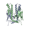
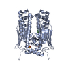
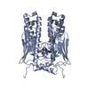
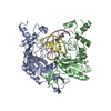
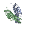
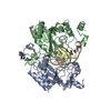
| Title | THE SUBSTRATE BINDING DOMAIN OF DNAK IN COMPLEX WITH A SUBSTRATE PEPTIDE, DETERMINED FROM TYPE 1 SELENOMETHIONYL CRYSTALS | ||||||
 Components Components |
| ||||||
 Keywords Keywords | COMPLEX (MOLECULAR CHAPERONE/PEPTIDE) / DNAK / HEAT SHOCK PROTEIN 70 KDA (HSP70) / COMPLEX (MOLECULAR CHAPERONE-PEPTIDE) / COMPLEX (MOLECULAR CHAPERONE-PEPTIDE) complex | ||||||
| Function / homology |  Function and homology information Function and homology informationstress response to copper ion / sigma factor antagonist activity / protein unfolding / cellular response to unfolded protein / heat shock protein binding / protein folding chaperone / inclusion body / ATP-dependent protein folding chaperone / ADP binding / : ...stress response to copper ion / sigma factor antagonist activity / protein unfolding / cellular response to unfolded protein / heat shock protein binding / protein folding chaperone / inclusion body / ATP-dependent protein folding chaperone / ADP binding / : / protein refolding / response to heat / protein-folding chaperone binding / protein folding / protein-containing complex assembly / DNA replication / ATP hydrolysis activity / protein-containing complex / zinc ion binding / ATP binding / membrane / plasma membrane / cytosol / cytoplasm Similarity search - Function | ||||||
| Biological species |  | ||||||
| Method |  X-RAY DIFFRACTION / Resolution: 2 Å X-RAY DIFFRACTION / Resolution: 2 Å | ||||||
 Authors Authors | Zhu, X. / Zhao, X. / Burkholder, W.F. / Gragerov, A. / Ogata, C.M. / Gottesman, M.E. / Hendrickson, W.A. | ||||||
 Citation Citation | Journal: Science / Year: 1996 Title: Structural analysis of substrate binding by the molecular chaperone DnaK. Authors: Zhu, X. / Zhao, X. / Burkholder, W.F. / Gragerov, A. / Ogata, C.M. / Gottesman, M.E. / Hendrickson, W.A. | ||||||
| History |
|
- Structure visualization
| Structure viewer | Molecule: MolmilJmol/JSmol |
|---|
- Downloads & links
-Download
| PDBx/mmCIF format | 1dkx.cif.gz | 60 KB | Display | PDBx/mmCIF format |
|---|---|---|---|---|
| PDB format | pdb1dkx.ent.gz | 44.1 KB | Display | PDB format |
| PDBx/mmJSON format | 1dkx.json.gz | Tree view | PDBx/mmJSON format | |
| Others |  Other downloads Other downloads |
-Validation report
| Arichive directory | https://data.pdbj.org/pub/pdb/validation_reports/dk/1dkxftp://data.pdbj.org/pub/pdb/validation_reports/dk/1dkx | HTTPS FTP |
|---|
-Related structure data


-Links
 PDBj
PDBj
- Assembly
| Deposited unit | 
| ||||||||
|---|---|---|---|---|---|---|---|---|---|
| 1 | 
| ||||||||
| 2 | 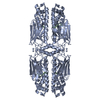
| ||||||||
| Unit cell |
|
-Components
| #1: Protein | Mass: 23806.752 Da / Num. of mol.: 1 / Source method: isolated from a natural source / Source: (natural) |
|---|---|
| #2: Protein/peptide | Mass: 786.941 Da / Num. of mol.: 1 Source method: isolated from a genetically manipulated source |
| #3: Water | ChemComp-HOH /  Mass: 18.015 Da / Num. of mol.: 216 / Source method: isolated from a natural source / Formula: H2O Mass: 18.015 Da / Num. of mol.: 216 / Source method: isolated from a natural source / Formula: H2O |
-Experimental details
-Experiment
| Experiment | Method: X-RAY DIFFRACTION |
|---|
- Sample preparation
| Crystal | Density Matthews: 2.08 Å3/Da / Density % sol: 43 % | |||||||||||||||
|---|---|---|---|---|---|---|---|---|---|---|---|---|---|---|---|---|
| Crystal grow | *PLUS pH: 7 / Method: vapor diffusion, hanging drop / Details: or dialysis | |||||||||||||||
| Components of the solutions | *PLUS
|
-Data collection
| Radiation | Scattering type: x-ray |
|---|---|
| Radiation wavelength | Relative weight: 1 |
| Reflection | Resolution: 2→20 Å / Num. obs: 13579 / % possible obs: 94.9 % / Observed criterion σ(I): 2 / Redundancy: 37 % |
| Reflection | *PLUS Num. measured all: 59575 / Rmerge(I) obs: 0.052 |
| Reflection shell | *PLUS Highest resolution: 2 Å / Lowest resolution: 2.1 Å / % possible obs: 84.6 % / Rmerge(I) obs: 0.185 |
- Processing
| Software |
| ||||||||||||||||||||||||||||||||||||||||||||||||||||||||||||
|---|---|---|---|---|---|---|---|---|---|---|---|---|---|---|---|---|---|---|---|---|---|---|---|---|---|---|---|---|---|---|---|---|---|---|---|---|---|---|---|---|---|---|---|---|---|---|---|---|---|---|---|---|---|---|---|---|---|---|---|---|---|
| Refinement | Resolution: 2→5 Å / σ(F): 2
| ||||||||||||||||||||||||||||||||||||||||||||||||||||||||||||
| Displacement parameters | Biso mean: 23.26 Å2 | ||||||||||||||||||||||||||||||||||||||||||||||||||||||||||||
| Refinement step | Cycle: LAST / Resolution: 2→5 Å
| ||||||||||||||||||||||||||||||||||||||||||||||||||||||||||||
| Refine LS restraints |
| ||||||||||||||||||||||||||||||||||||||||||||||||||||||||||||
| Refinement | *PLUS | ||||||||||||||||||||||||||||||||||||||||||||||||||||||||||||
| Solvent computation | *PLUS | ||||||||||||||||||||||||||||||||||||||||||||||||||||||||||||
| Displacement parameters | *PLUS | ||||||||||||||||||||||||||||||||||||||||||||||||||||||||||||
| Refine LS restraints | *PLUS
|
