- PDB-2j96: The E-configuration of alfa-Phycoerythrocyanin -
+
Open data
ID or keywords:
Loading...
-
Basic information
Entry
Database: PDB / ID: 2j96
Title
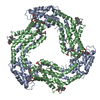
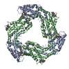
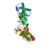
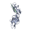
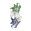
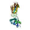
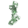
The E-configuration of alfa-Phycoerythrocyanin
Components
PHYCOERYTHROCYANIN ALPHA CHAIN
Keywords
PHOTOSYNTHESIS / ELECTRON TRANSPORT / Z- TO E-ISOMERIZATION / TRANSPORT / CHROMOPHORE / BILE PIGMENT / PHYCOBILISOME / LIGHT HARVESTING / PHYCOBILIPROTEINS
Mass: 17581.686 Da / Num. of mol.: 2 / Source method: isolated from a natural source Details: PHYCOVIOLOBILIN CHROMOPHORE ATTACHED TO CYS84 AND REFINED AS A WHOLE Source: (natural) MASTIGOCLADUS LAMINOSUS (bacteria) / References: UniProt: P00309
#2: Chemical
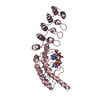
ChemComp-PVN / PHYCOVIOLOBILIN / Phycoviolobilin, bound form
Mass: 588.694 Da / Num. of mol.: 2 / Source method: obtained synthetically / Formula: C33H40N4O6
Resolution: 2.25→50 Å / Data cutoff high absF: 10000 / Isotropic thermal model: RESTRAINED / Cross valid method: THROUGHOUT / σ(F): 2 / Stereochemistry target values: MAXIMUM LIKELIHOOD Details: THE PVN CHROMOPHORE HAS BEEN ATTACHED TO CYS84 USING RESTRAINTS DERIVED FROM METHIONINE AND REFINED AS IF IT WOULD BE AN AMINO ACID
Rfactor
Num. reflection
% reflection
Selection details
Rfree
0.3086
898
4.5 %
RANDOM
Rwork
0.235
-
-
-
obs
0.235
17895
91.9 %
-
Solvent computation
Bsol: 21.1171 Å2 / ksol: 0.300406 e/Å3
Displacement parameters
Biso mean: 35.3 Å2
Baniso -1
Baniso -2
Baniso -3
1-
5.815 Å2
0 Å2
1.749 Å2
2-
-
-18.144 Å2
0 Å2
3-
-
-
12.329 Å2
Refine analyze
Free
Obs
Luzzati coordinate error
0.43 Å
0.29 Å
Luzzati d res low
-
5 Å
Luzzati sigma a
0.31 Å
0.21 Å
Refinement step
Cycle: LAST / Resolution: 2.25→50 Å
Protein
Nucleic acid
Ligand
Solvent
Total
Num. atoms
2478
0
86
326
2890
Refine LS restraints
Refine-ID
Type
Dev ideal
Dev ideal target
X-RAY DIFFRACTION
c_bond_d
0.01483
X-RAY DIFFRACTION
c_bond_d_na
X-RAY DIFFRACTION
c_bond_d_prot
X-RAY DIFFRACTION
c_angle_d
X-RAY DIFFRACTION
c_angle_d_na
X-RAY DIFFRACTION
c_angle_d_prot
X-RAY DIFFRACTION
c_angle_deg
1.68283
X-RAY DIFFRACTION
c_angle_deg_na
X-RAY DIFFRACTION
c_angle_deg_prot
X-RAY DIFFRACTION
c_dihedral_angle_d
21
X-RAY DIFFRACTION
c_dihedral_angle_d_na
X-RAY DIFFRACTION
c_dihedral_angle_d_prot
X-RAY DIFFRACTION
c_improper_angle_d
2.14
X-RAY DIFFRACTION
c_improper_angle_d_na
X-RAY DIFFRACTION
c_improper_angle_d_prot
X-RAY DIFFRACTION
c_mcbond_it
1.58
1.5
X-RAY DIFFRACTION
c_mcangle_it
2.67
2
X-RAY DIFFRACTION
c_scbond_it
2.01
2
X-RAY DIFFRACTION
c_scangle_it
2.95
2.5
LS refinement shell
Resolution: 2.25→2.33 Å / Total num. of bins used: 10
In the structure databanks used in Yorodumi, some data are registered as the other names, "COVID-19 virus" and "2019-nCoV". Here are the details of the virus and the list of structure data.
Jan 31, 2019. EMDB accession codes are about to change! (news from PDBe EMDB page)
EMDB accession codes are about to change! (news from PDBe EMDB page)
The allocation of 4 digits for EMDB accession codes will soon come to an end. Whilst these codes will remain in use, new EMDB accession codes will include an additional digit and will expand incrementally as the available range of codes is exhausted. The current 4-digit format prefixed with “EMD-” (i.e. EMD-XXXX) will advance to a 5-digit format (i.e. EMD-XXXXX), and so on. It is currently estimated that the 4-digit codes will be depleted around Spring 2019, at which point the 5-digit format will come into force.
The EM Navigator/Yorodumi systems omit the EMD- prefix.
Related info.:Q: What is EMD? / ID/Accession-code notation in Yorodumi/EM Navigator
Yorodumi is a browser for structure data from EMDB, PDB, SASBDB, etc.
This page is also the successor to EM Navigator detail page, and also detail information page/front-end page for Omokage search.
The word "yorodu" (or yorozu) is an old Japanese word meaning "ten thousand". "mi" (miru) is to see.
Related info.:EMDB / PDB / SASBDB / Comparison of 3 databanks / Yorodumi Search / Aug 31, 2016. New EM Navigator & Yorodumi / Yorodumi Papers / Jmol/JSmol / Function and homology information / Changes in new EM Navigator and Yorodumi
 Movie
Movie Controller


 Open data
 Basic information
 Components
 Keywords
 Function and homology information
 MASTIGOCLADUS LAMINOSUS (bacteria)
 X-RAY DIFFRACTION /
  Authors
 Citation
 Structure visualization
 Downloads & links
 Other downloads
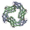


 PDBj
PDBj Assembly
 Mass: 588.694 Da / Num. of mol.: 2 / Source method: obtained synthetically / Formula: C33H40N4O6
 Mass: 18.015 Da / Num. of mol.: 326 / Source method: isolated from a natural source / Formula: H2O
 Sample preparation
 / Beamline: X13 / Wavelength: 1
  Processing