 Movie
Movie Controller

[English] 日本語
 Yorodumi
- PDB-5xcb: X-ray structure of domains D1 and D2 of Clostridium perfringens p... -
+ Open data

- Basic information
| Entry | Database: PDB / ID: 5xcb | ||||||
|---|---|---|---|---|---|---|---|
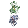
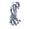
| Title | X-ray structure of domains D1 and D2 of Clostridium perfringens pili protein CppA | ||||||
 Components Components | Probable surface protein | ||||||
 Keywords Keywords | STRUCTURAL PROTEIN / pili protein | ||||||
| Function / homology | Gram-positive pilin backbone subunit 2, Cna-B-like domain / Gram-positive pilin backbone subunit 2, Cna-B-like domain / : / Fimbrial isopeptide formation D2 domain / Prealbumin-like fold domain / Prealbumin-like fold domain / Immunoglobulin-like fold / Probable surface protein Function and homology information Function and homology information | ||||||
| Biological species |  Clostridium perfringens str. 13 (bacteria) Clostridium perfringens str. 13 (bacteria) | ||||||
| Method |  X-RAY DIFFRACTION / MOLECULAR REPLACEMENT / Resolution: 2.14 Å X-RAY DIFFRACTION / MOLECULAR REPLACEMENT / Resolution: 2.14 Å | ||||||
 Authors Authors | Kamitori, S. / Tamai, E. | ||||||
| Funding support |  Japan, 1items Japan, 1items
| ||||||
 Citation Citation | Journal: Acta Crystallogr D Struct Biol / Year: 2019 Title: Structures of major pilins in Clostridium perfringens demonstrate dynamic conformational change. Authors: Tamai, E. / Katayama, S. / Sekiya, H. / Nariya, H. / Kamitori, S. | ||||||
| History |
|
- Structure visualization
| Structure viewer | Molecule: MolmilJmol/JSmol |
|---|
- Downloads & links
-Download
| PDBx/mmCIF format | 5xcb.cif.gz | 77.7 KB | Display | PDBx/mmCIF format |
|---|---|---|---|---|
| PDB format | pdb5xcb.ent.gz | 56.4 KB | Display | PDB format |
| PDBx/mmJSON format | 5xcb.json.gz | Tree view | PDBx/mmJSON format | |
| Others |  Other downloads Other downloads |
-Validation report
| Arichive directory | https://data.pdbj.org/pub/pdb/validation_reports/xc/5xcbftp://data.pdbj.org/pub/pdb/validation_reports/xc/5xcb | HTTPS FTP |
|---|
-Related structure data


-Links
 PDBj
PDBj- Assembly
| Deposited unit | 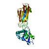
| ||||||||
|---|---|---|---|---|---|---|---|---|---|
| 1 |
| ||||||||
| Unit cell |
|
-Components
| #1: Protein | Mass: 36464.484 Da / Num. of mol.: 1 / Fragment: UNP residues 30-337 Source method: isolated from a genetically manipulated source Source: (gene. exp.) Clostridium perfringens str. 13 (bacteria)Strain: 13 / Gene: CPE0156 / Production host: |
|---|---|
| #2: Water | ChemComp-HOH /  Mass: 18.015 Da / Num. of mol.: 150 / Source method: isolated from a natural source / Formula: H2O Mass: 18.015 Da / Num. of mol.: 150 / Source method: isolated from a natural source / Formula: H2O |
| Has protein modification | Y |
-Experimental details
-Experiment
| Experiment | Method: X-RAY DIFFRACTION / Number of used crystals: 1 |
|---|
- Sample preparation
| Crystal | Density Matthews: 2.69 Å3/Da / Density % sol: 54.32 % |
|---|---|
| Crystal grow | Temperature: 293 K / Method: vapor diffusion, sitting drop / pH: 8.5 Details: 0.06M Magnesium chloride hexahydrate, 0.06M Calcium chloride dihydrate, 0.1M Tris (base), 0.1M BICINE, 20% v/v PEG 500* MME, 10 % w/v PEG 20000 |
-Data collection
| Diffraction | Mean temperature: 100 K |
|---|---|
| Diffraction source | Source: ROTATING ANODE / Type: RIGAKU MICROMAX-007 HF / Wavelength: 1.5418 Å |
| Detector | Type: RIGAKU RAXIS VII / Detector: IMAGE PLATE / Date: Nov 2, 2016 |
| Radiation | Protocol: SINGLE WAVELENGTH / Monochromatic (M) / Laue (L): M / Scattering type: x-ray |
| Radiation wavelength | Wavelength: 1.5418 Å / Relative weight: 1 |
| Reflection | Resolution: 2.14→18.76 Å / Num. obs: 22094 / % possible obs: 99.7 % / Redundancy: 6.5 % / Rmerge(I) obs: 0.052 / Net I/σ(I): 22.86 |
| Reflection shell | Resolution: 2.14→2.2 Å / Rmerge(I) obs: 0.487 / Mean I/σ(I) obs: 4.01 / % possible all: 99.8 |
- Processing
| Software |
| ||||||||||||||||||||||||||||||||||||||||||||||||||||||||||||||||||||||||||||||||||||||||||||||||||||||||||||||||||||||||||||||||||||||||||||||||||||||||||||||||||||||||||||||||||||||
|---|---|---|---|---|---|---|---|---|---|---|---|---|---|---|---|---|---|---|---|---|---|---|---|---|---|---|---|---|---|---|---|---|---|---|---|---|---|---|---|---|---|---|---|---|---|---|---|---|---|---|---|---|---|---|---|---|---|---|---|---|---|---|---|---|---|---|---|---|---|---|---|---|---|---|---|---|---|---|---|---|---|---|---|---|---|---|---|---|---|---|---|---|---|---|---|---|---|---|---|---|---|---|---|---|---|---|---|---|---|---|---|---|---|---|---|---|---|---|---|---|---|---|---|---|---|---|---|---|---|---|---|---|---|---|---|---|---|---|---|---|---|---|---|---|---|---|---|---|---|---|---|---|---|---|---|---|---|---|---|---|---|---|---|---|---|---|---|---|---|---|---|---|---|---|---|---|---|---|---|---|---|---|---|
| Refinement | Method to determine structure: MOLECULAR REPLACEMENT / Resolution: 2.14→18.76 Å / Cor.coef. Fo:Fc: 0.951 / Cor.coef. Fo:Fc free: 0.935 / SU B: 5.681 / SU ML: 0.146 / Cross valid method: THROUGHOUT / ESU R: 0.222 / ESU R Free: 0.19 / Details: HYDROGENS HAVE BEEN ADDED IN THE RIDING POSITIONS
| ||||||||||||||||||||||||||||||||||||||||||||||||||||||||||||||||||||||||||||||||||||||||||||||||||||||||||||||||||||||||||||||||||||||||||||||||||||||||||||||||||||||||||||||||||||||
| Solvent computation | Ion probe radii: 0.8 Å / Shrinkage radii: 0.8 Å / VDW probe radii: 1.2 Å | ||||||||||||||||||||||||||||||||||||||||||||||||||||||||||||||||||||||||||||||||||||||||||||||||||||||||||||||||||||||||||||||||||||||||||||||||||||||||||||||||||||||||||||||||||||||
| Displacement parameters | Biso mean: 47.056 Å2
| ||||||||||||||||||||||||||||||||||||||||||||||||||||||||||||||||||||||||||||||||||||||||||||||||||||||||||||||||||||||||||||||||||||||||||||||||||||||||||||||||||||||||||||||||||||||
| Refinement step | Cycle: 1 / Resolution: 2.14→18.76 Å
| ||||||||||||||||||||||||||||||||||||||||||||||||||||||||||||||||||||||||||||||||||||||||||||||||||||||||||||||||||||||||||||||||||||||||||||||||||||||||||||||||||||||||||||||||||||||
| Refine LS restraints |
|