 Movie
Movie Controller


+ Open data

- Basic information
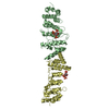
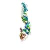
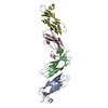
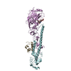
| Entry | Database: PDB / ID: 5xcc | ||||||
|---|---|---|---|---|---|---|---|
| Title | X-ray structure of Clostridium perfringens pili protein CppA | ||||||
 Components Components | Probable surface protein | ||||||
 Keywords Keywords | STRUCTURAL PROTEIN / pili protein | ||||||
| Function / homology | Gram-positive pilin backbone subunit 2, Cna-B-like domain / Gram-positive pilin backbone subunit 2, Cna-B-like domain / : / Fimbrial isopeptide formation D2 domain / Prealbumin-like fold domain / Prealbumin-like fold domain / Immunoglobulin-like fold / Probable surface protein Function and homology information Function and homology information | ||||||
| Biological species |  Clostridium perfringens str. 13 (bacteria) Clostridium perfringens str. 13 (bacteria) | ||||||
| Method |  X-RAY DIFFRACTION / SYNCHROTRON / MOLECULAR REPLACEMENT / Resolution: 2.48 Å X-RAY DIFFRACTION / SYNCHROTRON / MOLECULAR REPLACEMENT / Resolution: 2.48 Å | ||||||
 Authors Authors | Kamitori, S. / Tamai, E. | ||||||
| Funding support |  Japan, 1items Japan, 1items
| ||||||
 Citation Citation | Journal: Acta Crystallogr D Struct Biol / Year: 2019 Title: Structures of major pilins in Clostridium perfringens demonstrate dynamic conformational change. Authors: Tamai, E. / Katayama, S. / Sekiya, H. / Nariya, H. / Kamitori, S. | ||||||
| History |
|
- Structure visualization
| Structure viewer | Molecule: MolmilJmol/JSmol |
|---|
- Downloads & links
-Download
| PDBx/mmCIF format | 5xcc.cif.gz | 189.5 KB | Display | PDBx/mmCIF format |
|---|---|---|---|---|
| PDB format | pdb5xcc.ent.gz | 148.3 KB | Display | PDB format |
| PDBx/mmJSON format | 5xcc.json.gz | Tree view | PDBx/mmJSON format | |
| Others |  Other downloads Other downloads |
-Validation report
| Arichive directory | https://data.pdbj.org/pub/pdb/validation_reports/xc/5xccftp://data.pdbj.org/pub/pdb/validation_reports/xc/5xcc | HTTPS FTP |
|---|
-Related structure data
| Related structure data | 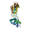 5xcbSC  6ixyC  6ixzC S: Starting model for refinement C: citing same article ( |
|---|---|
| Similar structure data |


-Links
 PDBj
PDBj- Assembly
| Deposited unit | 
| ||||||||
|---|---|---|---|---|---|---|---|---|---|
| 1 | 
| ||||||||
| 2 | 
| ||||||||
| Unit cell |
|
-Components
| #1: Protein | Mass: 52379.012 Da / Num. of mol.: 2 / Fragment: UNP residues 30-488 Source method: isolated from a genetically manipulated source Source: (gene. exp.) Clostridium perfringens str. 13 (bacteria)Strain: 13 / Gene: CPE0156 / Production host: #2: Water | ChemComp-HOH / |  Mass: 18.015 Da / Num. of mol.: 255 / Source method: isolated from a natural source / Formula: H2O Mass: 18.015 Da / Num. of mol.: 255 / Source method: isolated from a natural source / Formula: H2OHas protein modification | Y | |
|---|
-Experimental details
-Experiment
| Experiment | Method: X-RAY DIFFRACTION / Number of used crystals: 1 |
|---|
- Sample preparation
| Crystal | Density Matthews: 2.47 Å3/Da / Density % sol: 50.13 % |
|---|---|
| Crystal grow | Temperature: 293 K / Method: vapor diffusion, sitting drop / pH: 6.5 Details: 0.1M Sodium formate, 0.1M Ammonium acetate, 0.1M Sodium citrate tribasic dihydrate, 0.1M Sodium potassium tartrate tetrahydrate, 0.1M Sodium oxamate, 0.1M Imidazole, o.1M MES monohydrate ...Details: 0.1M Sodium formate, 0.1M Ammonium acetate, 0.1M Sodium citrate tribasic dihydrate, 0.1M Sodium potassium tartrate tetrahydrate, 0.1M Sodium oxamate, 0.1M Imidazole, o.1M MES monohydrate (acid), 20% v/v Ethylene glycol, 10% w/v PEG 8000 |
-Data collection
| Diffraction | Mean temperature: 100 K |
|---|---|
| Diffraction source | Source: SYNCHROTRON / Site: Photon Factory / Beamline: AR-NE3A / Wavelength: 1 Å |
| Detector | Type: ADSC QUANTUM 315 / Detector: CCD / Date: Feb 27, 2016 |
| Radiation | Protocol: SINGLE WAVELENGTH / Monochromatic (M) / Laue (L): M / Scattering type: x-ray |
| Radiation wavelength | Wavelength: 1 Å / Relative weight: 1 |
| Reflection | Resolution: 2.48→114.38 Å / Num. obs: 37465 / % possible obs: 99.4 % / Redundancy: 5.28 % / Rmerge(I) obs: 0.068 / Net I/σ(I): 16.08 |
| Reflection shell | Resolution: 2.48→2.54 Å / Rmerge(I) obs: 0.49 / Mean I/σ(I) obs: 3.71 / % possible all: 98.6 |
- Processing
| Software |
| ||||||||||||||||||||||||||||||||||||||||||||||||||||||||||||||||||||||||||||||||||||||||||||||||||||||||||||||||||||||||||||||||||||||||||||||||||||||||||||||||||||||||||||||||||||||
|---|---|---|---|---|---|---|---|---|---|---|---|---|---|---|---|---|---|---|---|---|---|---|---|---|---|---|---|---|---|---|---|---|---|---|---|---|---|---|---|---|---|---|---|---|---|---|---|---|---|---|---|---|---|---|---|---|---|---|---|---|---|---|---|---|---|---|---|---|---|---|---|---|---|---|---|---|---|---|---|---|---|---|---|---|---|---|---|---|---|---|---|---|---|---|---|---|---|---|---|---|---|---|---|---|---|---|---|---|---|---|---|---|---|---|---|---|---|---|---|---|---|---|---|---|---|---|---|---|---|---|---|---|---|---|---|---|---|---|---|---|---|---|---|---|---|---|---|---|---|---|---|---|---|---|---|---|---|---|---|---|---|---|---|---|---|---|---|---|---|---|---|---|---|---|---|---|---|---|---|---|---|---|---|
| Refinement | Method to determine structure: MOLECULAR REPLACEMENT Starting model: 5XCB Resolution: 2.48→114.38 Å / Cor.coef. Fo:Fc: 0.934 / Cor.coef. Fo:Fc free: 0.909 / SU B: 13.506 / SU ML: 0.284 / Cross valid method: THROUGHOUT / ESU R: 0.608 / ESU R Free: 0.311 / Details: HYDROGENS HAVE BEEN ADDED IN THE RIDING POSITIONS
| ||||||||||||||||||||||||||||||||||||||||||||||||||||||||||||||||||||||||||||||||||||||||||||||||||||||||||||||||||||||||||||||||||||||||||||||||||||||||||||||||||||||||||||||||||||||
| Solvent computation | Ion probe radii: 0.8 Å / Shrinkage radii: 0.8 Å / VDW probe radii: 1.2 Å | ||||||||||||||||||||||||||||||||||||||||||||||||||||||||||||||||||||||||||||||||||||||||||||||||||||||||||||||||||||||||||||||||||||||||||||||||||||||||||||||||||||||||||||||||||||||
| Displacement parameters | Biso mean: 46.56 Å2
| ||||||||||||||||||||||||||||||||||||||||||||||||||||||||||||||||||||||||||||||||||||||||||||||||||||||||||||||||||||||||||||||||||||||||||||||||||||||||||||||||||||||||||||||||||||||
| Refinement step | Cycle: 1 / Resolution: 2.48→114.38 Å
| ||||||||||||||||||||||||||||||||||||||||||||||||||||||||||||||||||||||||||||||||||||||||||||||||||||||||||||||||||||||||||||||||||||||||||||||||||||||||||||||||||||||||||||||||||||||
| Refine LS restraints |
|