 Movie
Movie Controller

[English] 日本語
 Yorodumi
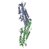
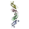
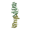
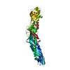
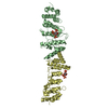
- PDB-1bp1: CRYSTAL STRUCTURE OF BPI, THE HUMAN BACTERICIDAL PERMEABILITY-INC... -
+ Open data

- Basic information
| Entry | Database: PDB / ID: 1bp1 | ||||||
|---|---|---|---|---|---|---|---|
| Title | CRYSTAL STRUCTURE OF BPI, THE HUMAN BACTERICIDAL PERMEABILITY-INCREASING PROTEIN | ||||||
 Components Components | BACTERICIDAL/PERMEABILITY-INCREASING PROTEIN | ||||||
 Keywords Keywords | BACTERICIDAL / PERMEABILITY-INCREASING / LIPID-BINDING / LIPOPOLYSACCHARIDE-BINDING / ANTIBIOTIC | ||||||
| Function / homology |  Function and homology information Function and homology informationnegative regulation of macrophage activation / Toll Like Receptor 4 (TLR4) Cascade / negative regulation of interleukin-8 production / Antimicrobial peptides / negative regulation of interleukin-6 production / negative regulation of tumor necrosis factor production / lipopolysaccharide-mediated signaling pathway / lipopolysaccharide binding / specific granule lumen / azurophil granule lumen ...negative regulation of macrophage activation / Toll Like Receptor 4 (TLR4) Cascade / negative regulation of interleukin-8 production / Antimicrobial peptides / negative regulation of interleukin-6 production / negative regulation of tumor necrosis factor production / lipopolysaccharide-mediated signaling pathway / lipopolysaccharide binding / specific granule lumen / azurophil granule lumen / defense response to Gram-negative bacterium / innate immune response / Neutrophil degranulation / : / extracellular exosome / extracellular region Similarity search - Function | ||||||
| Biological species |  Homo sapiens (human) Homo sapiens (human) | ||||||
| Method |  X-RAY DIFFRACTION / MIRAS / Resolution: 2.4 Å X-RAY DIFFRACTION / MIRAS / Resolution: 2.4 Å | ||||||
 Authors Authors | Beamer, L.J. / Carroll, S.F. / Eisenberg, D. | ||||||
 Citation Citation | Journal: Science / Year: 1997 Title: Crystal structure of human BPI and two bound phospholipids at 2.4 angstrom resolution. Authors: Beamer, L.J. / Carroll, S.F. / Eisenberg, D. #1: Journal: Infect.Agents Dis. / Year: 1995Title: Prospects for Use of Rbpi Fragments in the Treatment of Gram-Negative Bacterial Infections Authors: Elsbach, P. / Weiss, J. | ||||||
| History |
|
- Structure visualization
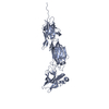
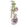
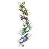
| Structure viewer | Molecule: MolmilJmol/JSmol |
|---|
- Downloads & links
-Download
| PDBx/mmCIF format | 1bp1.cif.gz | 103.9 KB | Display | PDBx/mmCIF format |
|---|---|---|---|---|
| PDB format | pdb1bp1.ent.gz | 79.2 KB | Display | PDB format |
| PDBx/mmJSON format | 1bp1.json.gz | Tree view | PDBx/mmJSON format | |
| Others |  Other downloads Other downloads |
-Validation report
| Arichive directory | https://data.pdbj.org/pub/pdb/validation_reports/bp/1bp1ftp://data.pdbj.org/pub/pdb/validation_reports/bp/1bp1 | HTTPS FTP |
|---|
-Related structure data
| Similar structure data |
|---|


-Links
 PDBj
PDBj


- Assembly
| Deposited unit | 
| ||||||||
|---|---|---|---|---|---|---|---|---|---|
| 1 |
| ||||||||
| Unit cell |
|
-Components
| #1: Protein | Mass: 50703.691 Da / Num. of mol.: 1 / Mutation: S351A Source method: isolated from a genetically manipulated source Source: (gene. exp.) Homo sapiens (human) / Cell: POLYMORPHONUCLEAR NEUTROPHILSCell line (production host): CHINESE HAMSTER OVARY CELLS (CHO) Cellular location (production host): PRIMARY GRANULES / Production host:   Cricetulus griseus (Chinese hamster) / References: UniProt: P17213 Cricetulus griseus (Chinese hamster) / References: UniProt: P17213 | ||||||
|---|---|---|---|---|---|---|---|
| #2: Chemical |   Mass: 790.145 Da / Num. of mol.: 2 / Source method: obtained synthetically / Formula: C44H88NO8P / Comment: phospholipid*YM Mass: 790.145 Da / Num. of mol.: 2 / Source method: obtained synthetically / Formula: C44H88NO8P / Comment: phospholipid*YM#3: Water | ChemComp-HOH / |  Mass: 18.015 Da / Num. of mol.: 48 / Source method: isolated from a natural source / Formula: H2O Mass: 18.015 Da / Num. of mol.: 48 / Source method: isolated from a natural source / Formula: H2OCompound details | BPI HAS TWO SIMILAR DOMAINS WHICH SHOW NO SIGNIFICAN | Has protein modification | Y | |
-Experimental details
-Experiment
| Experiment | Method: X-RAY DIFFRACTION / Number of used crystals: 2 |
|---|
- Sample preparation
| Crystal | Density Matthews: 2.52 Å3/Da / Density % sol: 51.2 % | |||||||||||||||||||||||||
|---|---|---|---|---|---|---|---|---|---|---|---|---|---|---|---|---|---|---|---|---|---|---|---|---|---|---|
| Crystal grow | pH: 6.8 Details: CRYSTALLIZED IN 12% PEG 8000, 0.2 M MG ACETATE, 0.1 NA CACODYLATE PH 6.8 | |||||||||||||||||||||||||
| Crystal grow | *PLUS Method: vapor diffusion, hanging drop | |||||||||||||||||||||||||
| Components of the solutions | *PLUS
|
-Data collection
| Diffraction | Mean temperature: 300 K |
|---|---|
| Diffraction source | Source: ROTATING ANODE / Type: RIGAKU RUH2R / Wavelength: 1.5418 |
| Detector | Type: RIGAKU / Detector: IMAGE PLATE / Date: Feb 1, 1994 / Details: MIRRORS |
| Radiation | Monochromator: NI FILTER / Monochromatic (M) / Laue (L): M / Scattering type: x-ray |
| Radiation wavelength | Wavelength: 1.5418 Å / Relative weight: 1 |
| Reflection | Resolution: 2.4→50 Å / Num. obs: 18908 / % possible obs: 92.7 % / Observed criterion σ(I): 0 / Biso Wilson estimate: 45 Å2 / Rsym value: 0.072 |
| Reflection shell | Resolution: 2.4→2.5 Å / Mean I/σ(I) obs: 2.6 / % possible all: 94.5 |
| Reflection | *PLUS Rmerge(I) obs: 0.072 |
| Reflection shell | *PLUS % possible obs: 94.5 % |
- Processing
| Software |
| ||||||||||||||||||||||||||||||||||||||||||||||||||||||||||||||||||||||||||||||||
|---|---|---|---|---|---|---|---|---|---|---|---|---|---|---|---|---|---|---|---|---|---|---|---|---|---|---|---|---|---|---|---|---|---|---|---|---|---|---|---|---|---|---|---|---|---|---|---|---|---|---|---|---|---|---|---|---|---|---|---|---|---|---|---|---|---|---|---|---|---|---|---|---|---|---|---|---|---|---|---|---|---|
| Refinement | Method to determine structure: MIRAS / Resolution: 2.4→50 Å / Rfactor Rfree error: 0.007 / Data cutoff high absF: 100000 / Data cutoff low absF: 0.1 / Isotropic thermal model: RESTRAINED / Cross valid method: THROUGHOUT / σ(F): 0 / Details: BULK SOLVENT MODEL USED
| ||||||||||||||||||||||||||||||||||||||||||||||||||||||||||||||||||||||||||||||||
| Displacement parameters | Biso mean: 45 Å2
| ||||||||||||||||||||||||||||||||||||||||||||||||||||||||||||||||||||||||||||||||
| Refine analyze |
| ||||||||||||||||||||||||||||||||||||||||||||||||||||||||||||||||||||||||||||||||
| Refinement step | Cycle: LAST / Resolution: 2.4→50 Å
| ||||||||||||||||||||||||||||||||||||||||||||||||||||||||||||||||||||||||||||||||
| Refine LS restraints |
| ||||||||||||||||||||||||||||||||||||||||||||||||||||||||||||||||||||||||||||||||
| LS refinement shell | Resolution: 2.4→2.55 Å / Rfactor Rfree error: 0.027 / Total num. of bins used: 6
| ||||||||||||||||||||||||||||||||||||||||||||||||||||||||||||||||||||||||||||||||
| Xplor file |
| ||||||||||||||||||||||||||||||||||||||||||||||||||||||||||||||||||||||||||||||||
| Software | *PLUS Name: X-PLOR / Version: 3.843 / Classification: refinement | ||||||||||||||||||||||||||||||||||||||||||||||||||||||||||||||||||||||||||||||||
| Refinement | *PLUS | ||||||||||||||||||||||||||||||||||||||||||||||||||||||||||||||||||||||||||||||||
| Solvent computation | *PLUS | ||||||||||||||||||||||||||||||||||||||||||||||||||||||||||||||||||||||||||||||||
| Displacement parameters | *PLUS | ||||||||||||||||||||||||||||||||||||||||||||||||||||||||||||||||||||||||||||||||
| Refine LS restraints | *PLUS
| ||||||||||||||||||||||||||||||||||||||||||||||||||||||||||||||||||||||||||||||||
| LS refinement shell | *PLUS Rfactor obs: 0.368 |
