 Movie
Movie Controller

[English] 日本語
 Yorodumi
- PDB-4g8l: Active state of intact sensor domain of human RNase L with 2-5A bound -
+ Open data

- Basic information
| Entry | Database: PDB / ID: 4g8l | ||||||
|---|---|---|---|---|---|---|---|
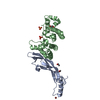
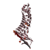
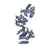
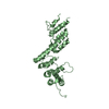
| Title | Active state of intact sensor domain of human RNase L with 2-5A bound | ||||||
 Components Components | 2-5A-dependent ribonuclease | ||||||
 Keywords Keywords | HYDROLASE / Ankyrin-repeat domain / single-stranded RNA | ||||||
| Function / homology |  Function and homology information Function and homology informationOAS antiviral response / Hydrolases; Acting on ester bonds; Endoribonucleases producing 5'-phosphomonoesters / negative regulation of viral genome replication / RNA nuclease activity / ribonucleoprotein complex binding / fat cell differentiation / RNA processing / RNA endonuclease activity / regulation of mRNA stability / positive regulation of D-glucose import across plasma membrane ...OAS antiviral response / Hydrolases; Acting on ester bonds; Endoribonucleases producing 5'-phosphomonoesters / negative regulation of viral genome replication / RNA nuclease activity / ribonucleoprotein complex binding / fat cell differentiation / RNA processing / RNA endonuclease activity / regulation of mRNA stability / positive regulation of D-glucose import across plasma membrane / nuclear matrix / mRNA processing / rRNA processing / Interferon alpha/beta signaling / defense response to virus / protein phosphorylation / protein kinase activity / rRNA binding / mitochondrial matrix / hydrolase activity / positive regulation of transcription by RNA polymerase II / RNA binding / zinc ion binding / ATP binding / identical protein binding / cytosol Similarity search - Function | ||||||
| Biological species |  Homo sapiens (human) Homo sapiens (human) | ||||||
| Method |  X-RAY DIFFRACTION / SYNCHROTRON / MOLECULAR REPLACEMENT / Resolution: 2.8 Å X-RAY DIFFRACTION / SYNCHROTRON / MOLECULAR REPLACEMENT / Resolution: 2.8 Å | ||||||
 Authors Authors | Han, Y. / Whitney, G. / Donovan, J. / Korennykh, A. | ||||||
 Citation Citation | Journal: Cell Rep / Year: 2012 Title: Innate Immune Messenger 2-5A Tethers Human RNase L into Active High-Order Complexes. Authors: Han, Y. / Whitney, G. / Donovan, J. / Korennykh, A. | ||||||
| History |
|
- Structure visualization
| Structure viewer | Molecule: MolmilJmol/JSmol |
|---|
- Downloads & links
-Download
| PDBx/mmCIF format | 4g8l.cif.gz | 473.1 KB | Display | PDBx/mmCIF format |
|---|---|---|---|---|
| PDB format | pdb4g8l.ent.gz | 392.2 KB | Display | PDB format |
| PDBx/mmJSON format | 4g8l.json.gz | Tree view | PDBx/mmJSON format | |
| Others |  Other downloads Other downloads |
-Validation report
| Arichive directory | https://data.pdbj.org/pub/pdb/validation_reports/g8/4g8lftp://data.pdbj.org/pub/pdb/validation_reports/g8/4g8l | HTTPS FTP |
|---|
-Related structure data
| Related structure data |  4g8kSC S: Starting model for refinement C: citing same article ( |
|---|---|
| Similar structure data |


-Links
 PDBj
PDBj

- Assembly
| Deposited unit | 
| |||||||||||||||||||||||||||||||||||||||||||||
|---|---|---|---|---|---|---|---|---|---|---|---|---|---|---|---|---|---|---|---|---|---|---|---|---|---|---|---|---|---|---|---|---|---|---|---|---|---|---|---|---|---|---|---|---|---|---|
| 1 | 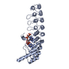
| |||||||||||||||||||||||||||||||||||||||||||||
| 2 | 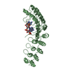
| |||||||||||||||||||||||||||||||||||||||||||||
| 3 | 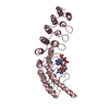
| |||||||||||||||||||||||||||||||||||||||||||||
| 4 | 
| |||||||||||||||||||||||||||||||||||||||||||||
| Unit cell |
| |||||||||||||||||||||||||||||||||||||||||||||
| Noncrystallographic symmetry (NCS) | NCS domain:
NCS domain segments:
NCS ensembles :
|
-Components
| #1: Protein | Mass: 37144.305 Da / Num. of mol.: 4 / Fragment: 2-5A-sensor domain (ANK domain) Source method: isolated from a genetically manipulated source Source: (gene. exp.) Homo sapiens (human) / Gene: RNASEL, RNS4 / Production host:  References: UniProt: Q05823, Hydrolases; Acting on ester bonds; Endoribonucleases producing 5'-phosphomonoesters #2: Chemical | ChemComp-25A /   Mass: 1005.633 Da / Num. of mol.: 4 / Source method: obtained synthetically / Formula: C30H38N15O19P3 Mass: 1005.633 Da / Num. of mol.: 4 / Source method: obtained synthetically / Formula: C30H38N15O19P3 |
|---|
-Experimental details
-Experiment
| Experiment | Method: X-RAY DIFFRACTION / Number of used crystals: 1 |
|---|
- Sample preparation
| Crystal | Density Matthews: 2.95 Å3/Da / Density % sol: 58.33 % |
|---|---|
| Crystal grow | Temperature: 293 K / Method: vapor diffusion / pH: 7 Details: 200 mM sodium acetate, 18% PEG 3350, 1.3M sodium citrate, pH 7.0, VAPOR DIFFUSION, temperature 293K |
-Data collection
| Diffraction | Mean temperature: 77 K | |||||||||||||||||||||||||||
|---|---|---|---|---|---|---|---|---|---|---|---|---|---|---|---|---|---|---|---|---|---|---|---|---|---|---|---|---|
| Diffraction source | Source: SYNCHROTRON / Site: NSLS  / Beamline: X29A / Wavelength: 1.072 Å / Beamline: X29A / Wavelength: 1.072 Å | |||||||||||||||||||||||||||
| Detector | Type: ADSC QUANTUM 315 / Detector: CCD / Date: Mar 11, 2012 | |||||||||||||||||||||||||||
| Radiation | Monochromator: Si 111 CHANNEL / Protocol: SINGLE WAVELENGTH / Monochromatic (M) / Laue (L): M / Scattering type: x-ray | |||||||||||||||||||||||||||
| Radiation wavelength | Wavelength: 1.072 Å / Relative weight: 1 | |||||||||||||||||||||||||||
| Reflection | Resolution: 2.8→39 Å / Num. obs: 42322 / % possible obs: 99.8 % / Observed criterion σ(F): 1.82 / Observed criterion σ(I): 1.82 | |||||||||||||||||||||||||||
| Reflection shell |
|
- Processing
| Software |
| |||||||||||||||||||||||||||||||||||||||||||||||||||||||||||||||||||||||||||||||||||||||||||||||||||||||||||||||||||||||||||||
|---|---|---|---|---|---|---|---|---|---|---|---|---|---|---|---|---|---|---|---|---|---|---|---|---|---|---|---|---|---|---|---|---|---|---|---|---|---|---|---|---|---|---|---|---|---|---|---|---|---|---|---|---|---|---|---|---|---|---|---|---|---|---|---|---|---|---|---|---|---|---|---|---|---|---|---|---|---|---|---|---|---|---|---|---|---|---|---|---|---|---|---|---|---|---|---|---|---|---|---|---|---|---|---|---|---|---|---|---|---|---|---|---|---|---|---|---|---|---|---|---|---|---|---|---|---|---|
| Refinement | Method to determine structure: MOLECULAR REPLACEMENT Starting model: PDB entry 4G8K Resolution: 2.8→38.75 Å / SU ML: 0.34 / σ(F): 1.99 / Phase error: 32.48 / Stereochemistry target values: ML
| |||||||||||||||||||||||||||||||||||||||||||||||||||||||||||||||||||||||||||||||||||||||||||||||||||||||||||||||||||||||||||||
| Solvent computation | Shrinkage radii: 0.9 Å / VDW probe radii: 1.11 Å / Solvent model: FLAT BULK SOLVENT MODEL / Bsol: 44.122 Å2 / ksol: 0.344 e/Å3 | |||||||||||||||||||||||||||||||||||||||||||||||||||||||||||||||||||||||||||||||||||||||||||||||||||||||||||||||||||||||||||||
| Displacement parameters |
| |||||||||||||||||||||||||||||||||||||||||||||||||||||||||||||||||||||||||||||||||||||||||||||||||||||||||||||||||||||||||||||
| Refinement step | Cycle: LAST / Resolution: 2.8→38.75 Å
| |||||||||||||||||||||||||||||||||||||||||||||||||||||||||||||||||||||||||||||||||||||||||||||||||||||||||||||||||||||||||||||
| Refine LS restraints |
| |||||||||||||||||||||||||||||||||||||||||||||||||||||||||||||||||||||||||||||||||||||||||||||||||||||||||||||||||||||||||||||
| Refine LS restraints NCS |
| |||||||||||||||||||||||||||||||||||||||||||||||||||||||||||||||||||||||||||||||||||||||||||||||||||||||||||||||||||||||||||||
| LS refinement shell |
| |||||||||||||||||||||||||||||||||||||||||||||||||||||||||||||||||||||||||||||||||||||||||||||||||||||||||||||||||||||||||||||
| Refinement TLS params. | Method: refined / Refine-ID: X-RAY DIFFRACTION
| |||||||||||||||||||||||||||||||||||||||||||||||||||||||||||||||||||||||||||||||||||||||||||||||||||||||||||||||||||||||||||||
| Refinement TLS group |
|
