- PDB-4ll6: Structure of Myo4p globular tail domain. -
+
Open data
ID or keywords:
Loading...
-
Basic information
Entry
Database: PDB / ID: 4ll6
Title
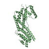
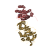
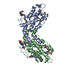
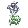
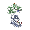
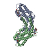
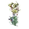
Structure of Myo4p globular tail domain.
Components
Myosin-4
Keywords
MOTOR PROTEIN / Myo4p / globular tail domain / mRNA localization
Function / homology
Function and homology information
Gap junction degradation / RHOT2 GTPase cycle / RHOT1 GTPase cycle / mating type switching / endoplasmic reticulum inheritance / RHOU GTPase cycle / cellular bud / vesicle transport along actin filament / cellular bud tip / intracellular mRNA localization ...Gap junction degradation / RHOT2 GTPase cycle / RHOT1 GTPase cycle / mating type switching / endoplasmic reticulum inheritance / RHOU GTPase cycle / cellular bud / vesicle transport along actin filament / cellular bud tip / intracellular mRNA localization / myosin complex / microfilament motor activity / filamentous actin / mRNA transport / actin filament organization / endocytosis / actin filament binding / actin cytoskeleton / calmodulin binding / mitochondrion / ATP binding / membrane / cytoplasm Similarity search - Function
Class V myosin, motor domain / Dilute domain / DIL domain / Dilute domain profile. / DIL / IQ calmodulin-binding motif / Myosin, N-terminal, SH3-like / Myosin N-terminal SH3-like domain profile. / Short calmodulin-binding motif containing conserved Ile and Gln residues. / IQ motif, EF-hand binding site ...Class V myosin, motor domain / Dilute domain / DIL domain / Dilute domain profile. / DIL / IQ calmodulin-binding motif / Myosin, N-terminal, SH3-like / Myosin N-terminal SH3-like domain profile. / Short calmodulin-binding motif containing conserved Ile and Gln residues. / IQ motif, EF-hand binding site / Myosin motor domain profile. / Myosin head, motor domain / Myosin head (motor domain) / Myosin. Large ATPases. / IQ motif profile. / Kinesin motor domain superfamily / P-loop containing nucleoside triphosphate hydrolase Similarity search - Domain/homology
In the structure databanks used in Yorodumi, some data are registered as the other names, "COVID-19 virus" and "2019-nCoV". Here are the details of the virus and the list of structure data.
Jan 31, 2019. EMDB accession codes are about to change! (news from PDBe EMDB page)
EMDB accession codes are about to change! (news from PDBe EMDB page)
The allocation of 4 digits for EMDB accession codes will soon come to an end. Whilst these codes will remain in use, new EMDB accession codes will include an additional digit and will expand incrementally as the available range of codes is exhausted. The current 4-digit format prefixed with “EMD-” (i.e. EMD-XXXX) will advance to a 5-digit format (i.e. EMD-XXXXX), and so on. It is currently estimated that the 4-digit codes will be depleted around Spring 2019, at which point the 5-digit format will come into force.
The EM Navigator/Yorodumi systems omit the EMD- prefix.
Related info.:Q: What is EMD? / ID/Accession-code notation in Yorodumi/EM Navigator
Yorodumi is a browser for structure data from EMDB, PDB, SASBDB, etc.
This page is also the successor to EM Navigator detail page, and also detail information page/front-end page for Omokage search.
The word "yorodu" (or yorozu) is an old Japanese word meaning "ten thousand". "mi" (miru) is to see.
Related info.:EMDB / PDB / SASBDB / Comparison of 3 databanks / Yorodumi Search / Aug 31, 2016. New EM Navigator & Yorodumi / Yorodumi Papers / Jmol/JSmol / Function and homology information / Changes in new EM Navigator and Yorodumi
 Movie
Movie Controller


 Open data
 Basic information
 Components
 Keywords
 Function and homology information
 X-RAY DIFFRACTION /
  Authors
 Citation
 Structure visualization
 Downloads & links
 Other downloads


 PDBj
PDBj


 Assembly
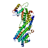


 Mass: 60.052 Da / Num. of mol.: 2 / Source method: obtained synthetically / Formula: C2H4O2
 Mass: 18.015 Da / Num. of mol.: 93 / Source method: isolated from a natural source / Formula: H2O
 Sample preparation
 / Beamline: X12C / Wavelength: 0.9798, 0.9801, 0.9574
 Processing