 Movie
Movie Controller

[English] 日本語
 Yorodumi
- PDB-4gsx: High resolution structure of dengue virus serotype 1 sE containin... -
+ Open data

- Basic information
| Entry | Database: PDB / ID: 4gsx | ||||||
|---|---|---|---|---|---|---|---|
| Title | High resolution structure of dengue virus serotype 1 sE containing stem | ||||||
 Components Components | Envelope protein E | ||||||
 Keywords Keywords | VIRAL PROTEIN / Viral Fusion Protein | ||||||
| Function / homology |  Function and homology information Function and homology informationflavivirin / host cell mitochondrion / symbiont-mediated suppression of host JAK-STAT cascade via inhibition of host TYK2 activity / symbiont-mediated suppression of host JAK-STAT cascade via inhibition of STAT2 activity / symbiont-mediated suppression of host cytoplasmic pattern recognition receptor signaling pathway via inhibition of MAVS activity / viral capsid / nucleoside-triphosphate phosphatase / double-stranded RNA binding / channel activity / monoatomic ion transmembrane transport ...flavivirin / host cell mitochondrion / symbiont-mediated suppression of host JAK-STAT cascade via inhibition of host TYK2 activity / symbiont-mediated suppression of host JAK-STAT cascade via inhibition of STAT2 activity / symbiont-mediated suppression of host cytoplasmic pattern recognition receptor signaling pathway via inhibition of MAVS activity / viral capsid / nucleoside-triphosphate phosphatase / double-stranded RNA binding / channel activity / monoatomic ion transmembrane transport / clathrin-dependent endocytosis of virus by host cell / mRNA (guanine-N7)-methyltransferase / methyltransferase cap1 / methyltransferase cap1 activity / mRNA 5'-cap (guanine-N7-)-methyltransferase activity / RNA helicase activity / protein dimerization activity / host cell perinuclear region of cytoplasm / host cell endoplasmic reticulum membrane / RNA helicase / symbiont-mediated suppression of host type I interferon-mediated signaling pathway / serine-type endopeptidase activity / symbiont-mediated activation of host autophagy / RNA-directed RNA polymerase / viral RNA genome replication / RNA-directed RNA polymerase activity / fusion of virus membrane with host endosome membrane / viral envelope / lipid binding / virion attachment to host cell / host cell nucleus / virion membrane / structural molecule activity / ATP hydrolysis activity / proteolysis / extracellular region / ATP binding / metal ion binding Similarity search - Function | ||||||
| Biological species |  Dengue virus 1 Dengue virus 1 | ||||||
| Method |  X-RAY DIFFRACTION / SYNCHROTRON / MOLECULAR REPLACEMENT / Resolution: 1.903 Å X-RAY DIFFRACTION / SYNCHROTRON / MOLECULAR REPLACEMENT / Resolution: 1.903 Å | ||||||
 Authors Authors | Klein, D.E. / Choi, J.L. / Harrison, S.C. | ||||||
 Citation Citation | Journal: J.Virol. / Year: 2013 Title: Structure of a dengue virus envelope protein late-stage fusion intermediate. Authors: Klein, D.E. / Choi, J.L. / Harrison, S.C. | ||||||
| History |
|
- Structure visualization
| Structure viewer | Molecule: MolmilJmol/JSmol |
|---|
- Downloads & links
-Download
| PDBx/mmCIF format | 4gsx.cif.gz | 181.4 KB | Display | PDBx/mmCIF format |
|---|---|---|---|---|
| PDB format | pdb4gsx.ent.gz | 140.4 KB | Display | PDB format |
| PDBx/mmJSON format | 4gsx.json.gz | Tree view | PDBx/mmJSON format | |
| Others |  Other downloads Other downloads |
-Validation report
| Arichive directory | https://data.pdbj.org/pub/pdb/validation_reports/gs/4gsxftp://data.pdbj.org/pub/pdb/validation_reports/gs/4gsx | HTTPS FTP |
|---|
-Related structure data
| Related structure data |  4gt0C  3g7tS S: Starting model for refinement C: citing same article ( |
|---|---|
| Similar structure data |


-Links
 PDBj
PDBj


- Assembly
| Deposited unit | 
| ||||||||||||
|---|---|---|---|---|---|---|---|---|---|---|---|---|---|
| 1 | 
| ||||||||||||
| 2 | 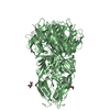
| ||||||||||||
| Unit cell |
| ||||||||||||
| Components on special symmetry positions |
|
-Components
| #1: Protein | Mass: 46796.188 Da / Num. of mol.: 2 / Fragment: UNP residues 281-691 / Mutation: W101H Source method: isolated from a genetically manipulated source Source: (gene. exp.) Dengue virus 1 / Strain: WP74 / Gene: Envelope Protein / Plasmid: pFastBac / Production host:  Trichoplusia ni (cabbage looper) / Strain (production host): Hi5 / References: UniProt: P17763 Trichoplusia ni (cabbage looper) / Strain (production host): Hi5 / References: UniProt: P17763#2: Sugar |   Type: D-saccharide, beta linking / Mass: 221.208 Da / Num. of mol.: 2 Type: D-saccharide, beta linking / Mass: 221.208 Da / Num. of mol.: 2Source method: isolated from a genetically manipulated source Formula: C8H15NO6 #3: Chemical | ChemComp-CD /   Mass: 112.411 Da / Num. of mol.: 4 / Source method: obtained synthetically / Formula: Cd Mass: 112.411 Da / Num. of mol.: 4 / Source method: obtained synthetically / Formula: Cd#4: Chemical | ChemComp-CL / |   Mass: 35.453 Da / Num. of mol.: 1 / Source method: obtained synthetically / Formula: Cl Mass: 35.453 Da / Num. of mol.: 1 / Source method: obtained synthetically / Formula: Cl#5: Water | ChemComp-HOH / |  Mass: 18.015 Da / Num. of mol.: 666 / Source method: isolated from a natural source / Formula: H2O Mass: 18.015 Da / Num. of mol.: 666 / Source method: isolated from a natural source / Formula: H2OHas protein modification | Y | |
|---|
-Experimental details
-Experiment
| Experiment | Method: X-RAY DIFFRACTION / Number of used crystals: 1 |
|---|
- Sample preparation
| Crystal | Density Matthews: 2.71 Å3/Da / Density % sol: 54.69 % |
|---|---|
| Crystal grow | Temperature: 293 K / Method: vapor diffusion, hanging drop / pH: 4.5 Details: pH 4.5, VAPOR DIFFUSION, HANGING DROP, temperature 293K |
-Data collection
| Diffraction | Mean temperature: 200 K |
|---|---|
| Diffraction source | Source: SYNCHROTRON / Site: APS  / Beamline: 24-ID-E / Wavelength: 0.979 Å / Beamline: 24-ID-E / Wavelength: 0.979 Å |
| Detector | Type: ADSC QUANTUM 315 / Detector: CCD / Date: Jun 11, 2011 / Details: mirrors |
| Radiation | Monochromator: Cryogenically-cooled single crystal / Protocol: SINGLE WAVELENGTH / Monochromatic (M) / Laue (L): M / Scattering type: x-ray |
| Radiation wavelength | Wavelength: 0.979 Å / Relative weight: 1 |
| Reflection twin | Type: merohedral / Operator: h,-h-k,-l / Fraction: 0.4 |
| Reflection | Resolution: 1.9→50 Å / Num. all: 76924 / Num. obs: 76924 / % possible obs: 99.1 % / Observed criterion σ(F): -3 / Observed criterion σ(I): -3 / Redundancy: 5.4 % / Rmerge(I) obs: 0.104 / Net I/σ(I): 14.2 |
| Reflection shell | Resolution: 1.9→1.97 Å / Redundancy: 5.1 % / Rmerge(I) obs: 0.445 / Mean I/σ(I) obs: 1.84 / Num. unique all: 7595 / % possible all: 99.3 |
- Processing
| Software |
| |||||||||||||||||||||||||||||||||||||||||||||||||||||||||||||||||||||||||||||||||||||||||||||||||||||||||||||||||||||||||||||||||||||||||||||||||||
|---|---|---|---|---|---|---|---|---|---|---|---|---|---|---|---|---|---|---|---|---|---|---|---|---|---|---|---|---|---|---|---|---|---|---|---|---|---|---|---|---|---|---|---|---|---|---|---|---|---|---|---|---|---|---|---|---|---|---|---|---|---|---|---|---|---|---|---|---|---|---|---|---|---|---|---|---|---|---|---|---|---|---|---|---|---|---|---|---|---|---|---|---|---|---|---|---|---|---|---|---|---|---|---|---|---|---|---|---|---|---|---|---|---|---|---|---|---|---|---|---|---|---|---|---|---|---|---|---|---|---|---|---|---|---|---|---|---|---|---|---|---|---|---|---|---|---|---|---|
| Refinement | Method to determine structure: MOLECULAR REPLACEMENT Starting model: PDB ENTRY 3G7T Resolution: 1.903→29.13 Å / σ(F): 1.4 / Phase error: 28.8 / Stereochemistry target values: TWIN_LSQ_F
| |||||||||||||||||||||||||||||||||||||||||||||||||||||||||||||||||||||||||||||||||||||||||||||||||||||||||||||||||||||||||||||||||||||||||||||||||||
| Solvent computation | Shrinkage radii: 0.9 Å / VDW probe radii: 1.11 Å / Solvent model: FLAT BULK SOLVENT MODEL | |||||||||||||||||||||||||||||||||||||||||||||||||||||||||||||||||||||||||||||||||||||||||||||||||||||||||||||||||||||||||||||||||||||||||||||||||||
| Refinement step | Cycle: LAST / Resolution: 1.903→29.13 Å
| |||||||||||||||||||||||||||||||||||||||||||||||||||||||||||||||||||||||||||||||||||||||||||||||||||||||||||||||||||||||||||||||||||||||||||||||||||
| Refine LS restraints |
| |||||||||||||||||||||||||||||||||||||||||||||||||||||||||||||||||||||||||||||||||||||||||||||||||||||||||||||||||||||||||||||||||||||||||||||||||||
| LS refinement shell |
|
