- PDB-4fg0: Structure of the St. Louis Encephalitis Virus envelope protein in... -
+
Open data
ID or keywords:
Loading...
-
Basic information
Entry
Database: PDB / ID: 4fg0
Title
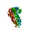
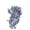
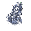
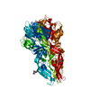
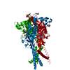
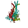
Structure of the St. Louis Encephalitis Virus envelope protein in the fusogenic trimer conformation.
Components
Polyprotein
Keywords
VIRAL PROTEIN / Viral envelope proteins / structural genomics / fusion peptide / antibody epitopes / Flavivirus / St. Louis Encephalitis Virus / NIAID / National Institute of Allergy and Infectious Diseases / Center for Structural Genomics of Infectious Diseases / CSGID
Function / homology
Function and homology information
flavivirin / symbiont-mediated suppression of host JAK-STAT cascade via inhibition of STAT2 activity / symbiont-mediated suppression of host JAK-STAT cascade via inhibition of STAT1 activity / viral capsid / nucleoside-triphosphate phosphatase / double-stranded RNA binding / clathrin-dependent endocytosis of virus by host cell / mRNA (guanine-N7)-methyltransferase / methyltransferase cap1 / methyltransferase cap1 activity ...flavivirin / symbiont-mediated suppression of host JAK-STAT cascade via inhibition of STAT2 activity / symbiont-mediated suppression of host JAK-STAT cascade via inhibition of STAT1 activity / viral capsid / nucleoside-triphosphate phosphatase / double-stranded RNA binding / clathrin-dependent endocytosis of virus by host cell / mRNA (guanine-N7)-methyltransferase / methyltransferase cap1 / methyltransferase cap1 activity / mRNA 5'-cap (guanine-N7-)-methyltransferase activity / RNA helicase activity / protein dimerization activity / symbiont-mediated suppression of host innate immune response / host cell perinuclear region of cytoplasm / host cell endoplasmic reticulum membrane / RNA helicase / symbiont-mediated suppression of host type I interferon-mediated signaling pathway / serine-type endopeptidase activity / RNA-directed RNA polymerase / viral RNA genome replication / RNA-directed RNA polymerase activity / fusion of virus membrane with host endosome membrane / viral envelope / symbiont entry into host cell / virion attachment to host cell / host cell nucleus / virion membrane / structural molecule activity / ATP hydrolysis activity / proteolysis / extracellular region / ATP binding / membrane / metal ion binding Similarity search - Function
Viral Envelope Glycoprotein, domain 2 / Viral Envelope Glycoprotein; domain 3 / Viral Envelope Glycoprotein, domain 3 / Tick-borne Encephalitis virus Glycoprotein, domain 1 / Viral Envelope Glycoprotein; domain 2 / Tick-borne Encephalitis virus Glycoprotein; domain 1 / Immunoglobulin-like - #350 / Flavivirus capsid protein C superfamily / Flavivirus non-structural protein NS2B / Genome polyprotein, Flavivirus ...Viral Envelope Glycoprotein, domain 2 / Viral Envelope Glycoprotein; domain 3 / Viral Envelope Glycoprotein, domain 3 / Tick-borne Encephalitis virus Glycoprotein, domain 1 / Viral Envelope Glycoprotein; domain 2 / Tick-borne Encephalitis virus Glycoprotein; domain 1 / Immunoglobulin-like - #350 / Flavivirus capsid protein C superfamily / Flavivirus non-structural protein NS2B / Genome polyprotein, Flavivirus / : / Flavivirus non-structural protein NS4A / Flavivirus non-structural protein NS2B / Flavivirus non-structural protein NS4B / mRNA cap 0/1 methyltransferase / Flavivirus non-structural protein NS4B / Flavivirus non-structural protein NS4A / Flavivirus NS2B domain profile. / mRNA cap 0 and cap 1 methyltransferase (EC 2.1.1.56 and EC 2.1.1.57) domain profile. / Flavivirus non-structural protein NS2A / Flavivirus non-structural protein NS2A / Flavivirus NS3, petidase S7 / Peptidase S7, Flavivirus NS3 serine protease / Flavivirus NS3 protease (NS3pro) domain profile. / RNA-directed RNA polymerase, thumb domain, Flavivirus / Flavivirus RNA-directed RNA polymerase, thumb domain / RNA-directed RNA polymerase, flavivirus / Flavivirus RNA-directed RNA polymerase, fingers and palm domains / Flavivirus capsid protein C / Flavivirus capsid protein C / Flavivirus non-structural Protein NS1 / Flavivirus non-structural protein NS1 / Envelope glycoprotein M superfamily, flavivirus / Envelope glycoprotein M, flavivirus / Flavivirus polyprotein propeptide superfamily / Flavivirus envelope glycoprotein M / Flavivirus polyprotein propeptide / Flavivirus polyprotein propeptide / : / Flavivirus NS3 helicase, C-terminal helical domain / Flavivirus envelope glycoprotein E, Stem/Anchor domain superfamily / Flavivirus envelope glycoprotein E, stem/anchor domain / Flavivirus envelope glycoprotein E, Stem/Anchor domain / Flaviviral glycoprotein E, central domain, subdomain 1 / Flaviviral glycoprotein E, central domain, subdomain 2 / Flavivirus glycoprotein E, immunoglobulin-like domain / Flavivirus glycoprotein, immunoglobulin-like domain / Flavivirus glycoprotein central and dimerisation domain / Flavivirus glycoprotein, central and dimerisation domains / Ribosomal RNA methyltransferase, FtsJ domain / FtsJ-like methyltransferase / Flavivirus/Alphavirus glycoprotein, immunoglobulin-like domain superfamily / Flavivirus glycoprotein, central and dimerisation domain superfamily / Flaviviral glycoprotein E, dimerisation domain / DEAD box, Flavivirus / Flavivirus DEAD domain / Immunoglobulin E-set / helicase superfamily c-terminal domain / Superfamilies 1 and 2 helicase C-terminal domain profile. / Superfamilies 1 and 2 helicase ATP-binding type-1 domain profile. / DEAD-like helicases superfamily / Helicase, C-terminal / Helicase superfamily 1/2, ATP-binding domain / RNA-directed RNA polymerase, catalytic domain / RdRp of positive ssRNA viruses catalytic domain profile. / S-adenosyl-L-methionine-dependent methyltransferase superfamily / Peptidase S1, PA clan / DNA/RNA polymerase superfamily / Immunoglobulin-like / Sandwich / P-loop containing nucleoside triphosphate hydrolase / 2-Layer Sandwich / Mainly Beta / Alpha Beta Similarity search - Domain/homology
Mass: 44613.996 Da / Num. of mol.: 1 Source method: isolated from a genetically manipulated source Details: refolded / Source: (gene. exp.) St. Louis encephalitis virus / Strain: St. Louis Encephalitis Virus, strain MS1-7 / Gene: Envelope protein E / Plasmid: pET21a(+) / Production host: Escherichia coli (E. coli) / Strain (production host): BL21(DE3)RIL / References: UniProt: Q9ENF3, UniProt: P09732*PLUS
Has protein modification
Y
-
Experimental details
-
Experiment
Experiment
Method: X-RAY DIFFRACTION / Number of used crystals: 1
-
Sample preparation
Crystal
Density Matthews: 5.22 Å3/Da / Density % sol: 76.44 %
Redundancy: 22.1 % / Av σ(I) over netI: 30.36 / Number: 191328 / Rmerge(I) obs: 0.11 / Χ2: 1.02 / D res high: 3.9 Å / D res low: 35 Å / Num. obs: 8657 / % possible obs: 100
Diffraction reflection shell
Highest resolution (Å)
Lowest resolution (Å)
% possible obs (%)
ID
Rmerge(I) obs
Chi squared
Redundancy
8.37
35
100
1
0.058
1.098
20.9
6.66
8.37
100
1
0.082
1.055
21.3
5.82
6.66
100
1
0.119
1.01
21.9
5.29
5.82
100
1
0.117
1.034
22.2
4.91
5.29
100
1
0.122
1.087
22.3
4.62
4.91
100
1
0.129
1.005
22.5
4.39
4.62
100
1
0.202
1.002
22.5
4.2
4.39
100
1
0.266
0.983
22.5
4.04
4.2
100
1
0.433
0.95
22.5
3.9
4.04
100
1
0.733
0.965
22.6
Reflection
Resolution: 3.899→35 Å / Num. obs: 8657 / % possible obs: 100 % / Redundancy: 22.1 % / Rmerge(I) obs: 0.11 / Χ2: 1.019 / Net I/σ(I): 30.36
Reflection shell
Diffraction-ID: 1 / % possible all: 100
Resolution (Å)
Redundancy (%)
Rmerge(I) obs
Mean I/σ(I) obs
Num. unique all
Χ2
3.9-4.04
22.6
0.733
6.21
880
0.965
4.04-4.2
22.5
0.433
833
0.95
4.2-4.39
22.5
0.266
857
0.983
4.39-4.62
22.5
0.202
847
1.002
4.62-4.91
22.5
0.129
865
1.005
4.91-5.29
22.3
0.122
860
1.087
5.29-5.82
22.2
0.117
856
1.034
5.82-6.66
21.9
0.119
872
1.01
6.66-8.37
21.3
0.082
870
1.055
8.37-35
20.9
0.058
917
1.098
-
Phasing
Phasing
Method: molecular replacement
Phasing MR
Highest resolution
Lowest resolution
Rotation
4.03 Å
37.81 Å
Translation
4.03 Å
37.81 Å
-
Processing
Software
Name
Version
Classification
NB
DENZO
datareduction
SCALEPACK
datascaling
PHASER
phasing
PHENIX
1.7.2_869
refinement
PDB_EXTRACT
3.11
dataextraction
HKL-3000
datacollection
HKL-2000
datareduction
Refinement
Method to determine structure: MOLECULAR REPLACEMENT / Resolution: 3.899→34.808 Å / Occupancy max: 1 / Occupancy min: 1 / FOM work R set: 0.8137 / SU ML: 1.01 / σ(F): 0 / Phase error: 25.27 / Stereochemistry target values: MLHL
Rfactor
Num. reflection
% reflection
Rfree
0.2666
847
9.9 %
Rwork
0.2218
-
-
obs
0.2262
8555
98.87 %
Solvent computation
Shrinkage radii: 0.98 Å / VDW probe radii: 1.2 Å / Solvent model: FLAT BULK SOLVENT MODEL / Bsol: 82.227 Å2 / ksol: 0.284 e/Å3
In the structure databanks used in Yorodumi, some data are registered as the other names, "COVID-19 virus" and "2019-nCoV". Here are the details of the virus and the list of structure data.
Jan 31, 2019. EMDB accession codes are about to change! (news from PDBe EMDB page)
EMDB accession codes are about to change! (news from PDBe EMDB page)
The allocation of 4 digits for EMDB accession codes will soon come to an end. Whilst these codes will remain in use, new EMDB accession codes will include an additional digit and will expand incrementally as the available range of codes is exhausted. The current 4-digit format prefixed with “EMD-” (i.e. EMD-XXXX) will advance to a 5-digit format (i.e. EMD-XXXXX), and so on. It is currently estimated that the 4-digit codes will be depleted around Spring 2019, at which point the 5-digit format will come into force.
The EM Navigator/Yorodumi systems omit the EMD- prefix.
Related info.:Q: What is EMD? / ID/Accession-code notation in Yorodumi/EM Navigator
Yorodumi is a browser for structure data from EMDB, PDB, SASBDB, etc.
This page is also the successor to EM Navigator detail page, and also detail information page/front-end page for Omokage search.
The word "yorodu" (or yorozu) is an old Japanese word meaning "ten thousand". "mi" (miru) is to see.
Related info.:EMDB / PDB / SASBDB / Comparison of 3 databanks / Yorodumi Search / Aug 31, 2016. New EM Navigator & Yorodumi / Yorodumi Papers / Jmol/JSmol / Function and homology information / Changes in new EM Navigator and Yorodumi
 Movie
Movie Controller

 Yorodumi
 Open data
 Basic information
 Components
 Keywords
 Function and homology information
 St. Louis encephalitis virus
 X-RAY DIFFRACTION /
  Authors
 Citation
 Structure visualization
 Downloads & links
 Other downloads


 PDBj
PDBj


 Assembly
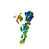

 Sample preparation
 / Beamline: 19-ID / Wavelength: 0.97935 Å
 Processing