 Movie
Movie Controller

[English] 日本語
 Yorodumi
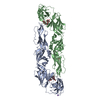
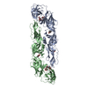
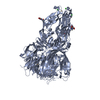
- PDB-1ok8: Crystal structure of the dengue 2 virus envelope glycoprotein in ... -
+ Open data

- Basic information
| Entry | Database: PDB / ID: 1ok8 | ||||||
|---|---|---|---|---|---|---|---|
| Title | Crystal structure of the dengue 2 virus envelope glycoprotein in the postfusion conformation | ||||||
 Components Components | MAJOR ENVELOPE PROTEIN E | ||||||
 Keywords Keywords | VIRAL PROTEIN / MEMBRANE FUSION / FLAVIVIRUS / FUSION PEPTIDE / LOW-PH CONFORMATIONAL CHANGE / CLASS 2 FUSION PROTEIN | ||||||
| Function / homology |  Function and homology information Function and homology informationhost cell nucleolus / flavivirin / host cell mitochondrion / symbiont-mediated suppression of host JAK-STAT cascade via inhibition of host TYK2 activity / symbiont-mediated suppression of host JAK-STAT cascade via inhibition of STAT2 activity / symbiont-mediated suppression of host cytoplasmic pattern recognition receptor signaling pathway via inhibition of MAVS activity / viral capsid / nucleoside-triphosphate phosphatase / double-stranded RNA binding / channel activity ...host cell nucleolus / flavivirin / host cell mitochondrion / symbiont-mediated suppression of host JAK-STAT cascade via inhibition of host TYK2 activity / symbiont-mediated suppression of host JAK-STAT cascade via inhibition of STAT2 activity / symbiont-mediated suppression of host cytoplasmic pattern recognition receptor signaling pathway via inhibition of MAVS activity / viral capsid / nucleoside-triphosphate phosphatase / double-stranded RNA binding / channel activity / monoatomic ion transmembrane transport / clathrin-dependent endocytosis of virus by host cell / mRNA (guanine-N7)-methyltransferase / molecular adaptor activity / methyltransferase cap1 / methyltransferase cap1 activity / mRNA 5'-cap (guanine-N7-)-methyltransferase activity / RNA helicase activity / protein dimerization activity / host cell perinuclear region of cytoplasm / host cell endoplasmic reticulum membrane / RNA helicase / symbiont-mediated suppression of host type I interferon-mediated signaling pathway / serine-type endopeptidase activity / symbiont-mediated activation of host autophagy / RNA-directed RNA polymerase / viral RNA genome replication / RNA-directed RNA polymerase activity / fusion of virus membrane with host endosome membrane / viral envelope / lipid binding / virion attachment to host cell / virion membrane / structural molecule activity / DNA-templated transcription / ATP hydrolysis activity / proteolysis / extracellular region / ATP binding / metal ion binding Similarity search - Function | ||||||
| Biological species |  DENGUE VIRUS TYPE 2 DENGUE VIRUS TYPE 2 | ||||||
| Method |  X-RAY DIFFRACTION / SYNCHROTRON / MOLECULAR REPLACEMENT / Resolution: 2 Å X-RAY DIFFRACTION / SYNCHROTRON / MOLECULAR REPLACEMENT / Resolution: 2 Å | ||||||
 Authors Authors | Modis, Y. / Harrison, S.C. | ||||||
 Citation Citation | Journal: Nature / Year: 2004 Title: Structure of the Dengue Virus Envelope Protein After Membrane Fusion Authors: Modis, Y. / Ogata, S. / Clements, D. / Harrison, S.C. #1: Journal: Proc.Natl.Acad.Sci.USA. / Year: 2003Title: A Ligand-Binding Pocket in the Dengue Virus Envelope Glycoprotein Authors: Modis, Y. / Ogata, S. / Clements, D. / Harrison, S.C. | ||||||
| History |
| ||||||
| Remark 650 | HELIX DETERMINATION METHOD: AUTHOR PROVIDED. | ||||||
| Remark 700 | SHEET THE SHEET STRUCTURE OF THIS MOLECULE IS BIFURCATED. IN ORDER TO REPRESENT THIS FEATURE IN ... SHEET THE SHEET STRUCTURE OF THIS MOLECULE IS BIFURCATED. IN ORDER TO REPRESENT THIS FEATURE IN THE SHEET RECORDS BELOW, TWO SHEETS ARE DEFINED. |
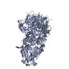
- Structure visualization
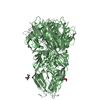
| Structure viewer | Molecule: MolmilJmol/JSmol |
|---|
- Downloads & links
-Download
| PDBx/mmCIF format | 1ok8.cif.gz | 93.4 KB | Display | PDBx/mmCIF format |
|---|---|---|---|---|
| PDB format | pdb1ok8.ent.gz | 70.3 KB | Display | PDB format |
| PDBx/mmJSON format | 1ok8.json.gz | Tree view | PDBx/mmJSON format | |
| Others |  Other downloads Other downloads |
-Validation report
| Arichive directory | https://data.pdbj.org/pub/pdb/validation_reports/ok/1ok8ftp://data.pdbj.org/pub/pdb/validation_reports/ok/1ok8 | HTTPS FTP |
|---|
-Related structure data
| Related structure data |  1oam S: Starting model for refinement |
|---|---|
| Similar structure data |


-Links
 PDBj
PDBj


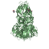
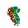
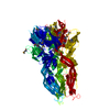
- Assembly
| Deposited unit | 
| |||||||||||||||
|---|---|---|---|---|---|---|---|---|---|---|---|---|---|---|---|---|
| 1 | 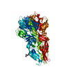
| |||||||||||||||
| Unit cell |
| |||||||||||||||
| Components on special symmetry positions |
|
-Components
| #1: Protein | Mass: 43819.391 Da / Num. of mol.: 1 / Fragment: SOLUBLE ECTODOMAIN, RESIDUES 281-674 Source method: isolated from a genetically manipulated source Source: (gene. exp.) DENGUE VIRUS TYPE 2 / Strain: PR159/S1 / Plasmid: PMTT / Cell line (production host): SCHNEIDER 2 / Production host:  |
|---|---|
| #2: Sugar | ChemComp-NAG /   Type: D-saccharide, beta linking / Mass: 221.208 Da / Num. of mol.: 1 Type: D-saccharide, beta linking / Mass: 221.208 Da / Num. of mol.: 1Source method: isolated from a genetically manipulated source Formula: C8H15NO6 |
| #3: Chemical | ChemComp-CL /   Mass: 35.453 Da / Num. of mol.: 1 / Source method: obtained synthetically / Formula: Cl Mass: 35.453 Da / Num. of mol.: 1 / Source method: obtained synthetically / Formula: Cl |
| #4: Water | ChemComp-HOH /  Mass: 18.015 Da / Num. of mol.: 205 / Source method: isolated from a natural source / Formula: H2O Mass: 18.015 Da / Num. of mol.: 205 / Source method: isolated from a natural source / Formula: H2O |
| Has protein modification | Y |
| Sequence details | RESIDUES 100-108 FORM THE GLYCINE-RICH, HYDROPHOBIC FUSION PEPTIDE (ALLISON ET AL., J.VIROL. 75, ...RESIDUES 100-108 FORM THE GLYCINE-RICH, HYDROPHOBI |
-Experimental details
-Experiment
| Experiment | Method: X-RAY DIFFRACTION / Number of used crystals: 1 |
|---|
- Sample preparation
| Crystal | Density Matthews: 2.445 Å3/Da / Density % sol: 49 % Description: THE THREE DOMAINS OF 1OAM WERE PLACED SEQUENTIALLY IN THE FOLLOWING ORDER: DOMAIN II, DOMAIN I, DOMAIN III. | ||||||||||||||||||||||||||||||||||||||||||||||||||||||||
|---|---|---|---|---|---|---|---|---|---|---|---|---|---|---|---|---|---|---|---|---|---|---|---|---|---|---|---|---|---|---|---|---|---|---|---|---|---|---|---|---|---|---|---|---|---|---|---|---|---|---|---|---|---|---|---|---|---|
| Crystal grow | pH: 7 / Details: 20-30% PEG 400, 0.08 M NACL, 0.1 M MOPS PH 7-8. | ||||||||||||||||||||||||||||||||||||||||||||||||||||||||
| Crystal grow | *PLUS Temperature: 20 ℃ / Method: vapor diffusion, hanging drop / PH range low: 8 / PH range high: 7 | ||||||||||||||||||||||||||||||||||||||||||||||||||||||||
| Components of the solutions | *PLUS
|
-Data collection
| Diffraction | Mean temperature: 100 K |
|---|---|
| Diffraction source | Source: SYNCHROTRON / Site: APS  / Beamline: 14-BM-C / Wavelength: 0.9 / Beamline: 14-BM-C / Wavelength: 0.9 |
| Detector | Type: ADSC CCD / Detector: CCD / Date: Mar 15, 2003 |
| Radiation | Protocol: SINGLE WAVELENGTH / Monochromatic (M) / Laue (L): M / Scattering type: x-ray |
| Radiation wavelength | Wavelength: 0.9 Å / Relative weight: 1 |
| Reflection | Resolution: 2→50 Å / Num. obs: 28677 / % possible obs: 95 % / Redundancy: 7 % / Biso Wilson estimate: 11.3 Å2 / Rmerge(I) obs: 0.058 / Net I/σ(I): 25 |
| Reflection shell | Resolution: 2→2.07 Å / Redundancy: 1.2 % / Rmerge(I) obs: 0.398 / Mean I/σ(I) obs: 2 / % possible all: 75 |
| Reflection | *PLUS Num. obs: 27450 / % possible obs: 95 % / Rmerge(I) obs: 0.058 |
| Reflection shell | *PLUS % possible obs: 75 % / Rmerge(I) obs: 0.398 / Mean I/σ(I) obs: 2 |
- Processing
| Software |
| ||||||||||||||||||||||||||||||||||||||||||||||||||||||||||||||||||||||||||||||||
|---|---|---|---|---|---|---|---|---|---|---|---|---|---|---|---|---|---|---|---|---|---|---|---|---|---|---|---|---|---|---|---|---|---|---|---|---|---|---|---|---|---|---|---|---|---|---|---|---|---|---|---|---|---|---|---|---|---|---|---|---|---|---|---|---|---|---|---|---|---|---|---|---|---|---|---|---|---|---|---|---|---|
| Refinement | Method to determine structure: MOLECULAR REPLACEMENT Starting model: PDB ENTRY 1OAM 1oam Resolution: 2→29.29 Å / Rfactor Rfree error: 0.007 / Data cutoff high absF: 441803.91 / Data cutoff low absF: 0 / Isotropic thermal model: RESTRAINED / Cross valid method: THROUGHOUT / σ(F): 0
| ||||||||||||||||||||||||||||||||||||||||||||||||||||||||||||||||||||||||||||||||
| Solvent computation | Solvent model: FLAT MODEL / Bsol: 47.9914 Å2 / ksol: 0.355448 e/Å3 | ||||||||||||||||||||||||||||||||||||||||||||||||||||||||||||||||||||||||||||||||
| Displacement parameters | Biso mean: 30.1 Å2
| ||||||||||||||||||||||||||||||||||||||||||||||||||||||||||||||||||||||||||||||||
| Refine analyze |
| ||||||||||||||||||||||||||||||||||||||||||||||||||||||||||||||||||||||||||||||||
| Refinement step | Cycle: LAST / Resolution: 2→29.29 Å
| ||||||||||||||||||||||||||||||||||||||||||||||||||||||||||||||||||||||||||||||||
| Refine LS restraints |
| ||||||||||||||||||||||||||||||||||||||||||||||||||||||||||||||||||||||||||||||||
| LS refinement shell | Resolution: 2→2.13 Å / Rfactor Rfree error: 0.023 / Total num. of bins used: 6
| ||||||||||||||||||||||||||||||||||||||||||||||||||||||||||||||||||||||||||||||||
| Xplor file |
| ||||||||||||||||||||||||||||||||||||||||||||||||||||||||||||||||||||||||||||||||
| Refinement | *PLUS Highest resolution: 2 Å / Lowest resolution: 30 Å / % reflection Rfree: 5 % / Rfactor Rfree: 0.2671 / Rfactor Rwork: 0.2213 | ||||||||||||||||||||||||||||||||||||||||||||||||||||||||||||||||||||||||||||||||
| Solvent computation | *PLUS | ||||||||||||||||||||||||||||||||||||||||||||||||||||||||||||||||||||||||||||||||
| Displacement parameters | *PLUS | ||||||||||||||||||||||||||||||||||||||||||||||||||||||||||||||||||||||||||||||||
| Refine LS restraints | *PLUS
|
