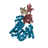
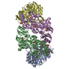
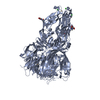
[English] 日本語
Yorodumi
- PDB-4gt0: Structure of dengue virus serotype 1 sE containing stem to residue 421 -

+
Open data


ID or keywords:

Loading...

-
Basic information

Entry
Database: PDB / ID: 4gt0
TitleStructure of dengue virus serotype 1 sE containing stem to residue 421
ComponentsEnvelope protein E
KeywordsVIRAL PROTEIN / Viral fusion protein
Function / homology
Function and homology information


symbiont-mediated suppression of host JAK-STAT cascade via inhibition of host TYK2 activity / flavivirin / host cell mitochondrion / symbiont-mediated suppression of host JAK-STAT cascade via inhibition of STAT2 activity / symbiont-mediated suppression of host cytoplasmic pattern recognition receptor signaling pathway via inhibition of MAVS activity / viral capsid / : / nucleoside-triphosphate phosphatase / double-stranded RNA binding / protein complex oligomerization ...symbiont-mediated suppression of host JAK-STAT cascade via inhibition of host TYK2 activity / flavivirin / host cell mitochondrion / symbiont-mediated suppression of host JAK-STAT cascade via inhibition of STAT2 activity / symbiont-mediated suppression of host cytoplasmic pattern recognition receptor signaling pathway via inhibition of MAVS activity / viral capsid / : / nucleoside-triphosphate phosphatase / double-stranded RNA binding / protein complex oligomerization / monoatomic ion channel activity / mRNA (guanine-N7)-methyltransferase / methyltransferase cap1 / clathrin-dependent endocytosis of virus by host cell / mRNA (nucleoside-2'-O-)-methyltransferase activity / mRNA 5'-cap (guanine-N7-)-methyltransferase activity / RNA helicase activity / host cell endoplasmic reticulum membrane / host cell perinuclear region of cytoplasm / protein dimerization activity / RNA helicase / induction by virus of host autophagy / RNA-directed RNA polymerase / viral RNA genome replication / RNA-dependent RNA polymerase activity / serine-type endopeptidase activity / fusion of virus membrane with host endosome membrane / viral envelope / symbiont-mediated suppression of host type I interferon-mediated signaling pathway / host cell nucleus / virion attachment to host cell / virion membrane / structural molecule activity / ATP hydrolysis activity / proteolysis / extracellular region / ATP binding / membrane / metal ion binding
Similarity search - Function
Viral Envelope Glycoprotein; domain 3 / Viral Envelope Glycoprotein, domain 3 / Viral Envelope Glycoprotein, domain 2 / Tick-borne Encephalitis virus Glycoprotein, domain 1 / Viral Envelope Glycoprotein; domain 2 / Tick-borne Encephalitis virus Glycoprotein; domain 1 / Immunoglobulin-like - #350 / : / Flavivirus envelope glycoprotein E, stem/anchor domain / RNA-directed RNA polymerase, thumb domain, Flavivirus ...Viral Envelope Glycoprotein; domain 3 / Viral Envelope Glycoprotein, domain 3 / Viral Envelope Glycoprotein, domain 2 / Tick-borne Encephalitis virus Glycoprotein, domain 1 / Viral Envelope Glycoprotein; domain 2 / Tick-borne Encephalitis virus Glycoprotein; domain 1 / Immunoglobulin-like - #350 / : / Flavivirus envelope glycoprotein E, stem/anchor domain / RNA-directed RNA polymerase, thumb domain, Flavivirus / Flavivirus RNA-directed RNA polymerase, thumb domain / Flavivirus capsid protein C superfamily / : / Flavivirus non-structural protein NS2B / Flavivirus NS3 helicase, C-terminal helical domain / Genome polyprotein, Flavivirus / Flavivirus non-structural protein NS4A / Flavivirus non-structural protein NS2B / Flavivirus capsid protein C / Flavivirus non-structural protein NS4B / mRNA cap 0/1 methyltransferase / Flavivirus capsid protein C / Flavivirus non-structural protein NS4B / Flavivirus non-structural protein NS4A / Flavivirus NS2B domain profile. / mRNA cap 0 and cap 1 methyltransferase (EC 2.1.1.56 and EC 2.1.1.57) domain profile. / Flavivirus non-structural protein NS2A / Flavivirus non-structural protein NS2A / Flavivirus NS3, petidase S7 / Peptidase S7, Flavivirus NS3 serine protease / Flavivirus NS3 protease (NS3pro) domain profile. / Envelope glycoprotein M, flavivirus / Flavivirus envelope glycoprotein M / RNA-directed RNA polymerase, flavivirus / Flavivirus RNA-directed RNA polymerase, fingers and palm domains / Flavivirus non-structural Protein NS1 / Flavivirus non-structural protein NS1 / Envelope glycoprotein M superfamily, flavivirus / Flavivirus polyprotein propeptide / Flavivirus polyprotein propeptide superfamily / Flavivirus polyprotein propeptide / Flaviviral glycoprotein E, central domain, subdomain 1 / Flaviviral glycoprotein E, central domain, subdomain 2 / Flavivirus envelope glycoprotein E, Stem/Anchor domain / Flavivirus glycoprotein E, immunoglobulin-like domain / Flavivirus envelope glycoprotein E, Stem/Anchor domain superfamily / Flavivirus glycoprotein, immunoglobulin-like domain / Flavivirus glycoprotein central and dimerisation domain / Flavivirus glycoprotein, central and dimerisation domains / Ribosomal RNA methyltransferase, FtsJ domain / FtsJ-like methyltransferase / Flavivirus/Alphavirus glycoprotein, immunoglobulin-like domain superfamily / Flavivirus glycoprotein, central and dimerisation domain superfamily / Flaviviral glycoprotein E, dimerisation domain / DEAD box, Flavivirus / Flavivirus DEAD domain / helicase superfamily c-terminal domain / Immunoglobulin E-set / Superfamilies 1 and 2 helicase C-terminal domain profile. / Superfamilies 1 and 2 helicase ATP-binding type-1 domain profile. / DEAD-like helicases superfamily / Helicase, C-terminal / Helicase superfamily 1/2, ATP-binding domain / RNA-directed RNA polymerase, catalytic domain / RdRp of positive ssRNA viruses catalytic domain profile. / S-adenosyl-L-methionine-dependent methyltransferase superfamily / Peptidase S1, PA clan, chymotrypsin-like fold / Peptidase S1, PA clan / DNA/RNA polymerase superfamily / Immunoglobulin-like / Sandwich / P-loop containing nucleoside triphosphate hydrolase / 2-Layer Sandwich / Mainly Beta / Alpha Beta
Similarity search - Domain/homology
: / Genome polyprotein
Similarity search - Component
Biological speciesDengue virus 1
MethodX-RAY DIFFRACTION / SYNCHROTRON / MOLECULAR REPLACEMENT / Resolution: 2.57 Å
AuthorsKlein, D.E. / Choi, J.L. / Harrison, S.C.
CitationJournal: J.Virol. / Year: 2013
Title: Structure of a dengue virus envelope protein late-stage fusion intermediate.
Authors: Klein, D.E. / Choi, J.L. / Harrison, S.C.
History
DepositionAug 28, 2012Deposition site: RCSB / Processing site: RCSB
Revision 1.0Dec 19, 2012Provider: repository / Type: Initial release
Revision 1.1Jan 30, 2013Group: Derived calculations
Revision 1.2Feb 6, 2013Group: Database references
Revision 1.3Nov 15, 2017Group: Refinement description / Category: software
Revision 1.4Jul 29, 2020Group: Data collection / Database references ...Data collection / Database references / Derived calculations / Structure summary
Category: chem_comp / entity ...chem_comp / entity / pdbx_chem_comp_identifier / pdbx_entity_nonpoly / pdbx_struct_conn_angle / struct_conn / struct_conn_type / struct_ref_seq_dif / struct_site / struct_site_gen
Item: _chem_comp.name / _chem_comp.type ..._chem_comp.name / _chem_comp.type / _entity.pdbx_description / _pdbx_entity_nonpoly.name / _pdbx_struct_conn_angle.ptnr1_auth_asym_id / _pdbx_struct_conn_angle.ptnr1_auth_comp_id / _pdbx_struct_conn_angle.ptnr1_auth_seq_id / _pdbx_struct_conn_angle.ptnr1_label_asym_id / _pdbx_struct_conn_angle.ptnr1_label_atom_id / _pdbx_struct_conn_angle.ptnr1_label_comp_id / _pdbx_struct_conn_angle.ptnr1_label_seq_id / _pdbx_struct_conn_angle.ptnr2_auth_asym_id / _pdbx_struct_conn_angle.ptnr2_auth_seq_id / _pdbx_struct_conn_angle.ptnr2_label_asym_id / _pdbx_struct_conn_angle.ptnr3_auth_asym_id / _pdbx_struct_conn_angle.ptnr3_auth_comp_id / _pdbx_struct_conn_angle.ptnr3_auth_seq_id / _pdbx_struct_conn_angle.ptnr3_label_asym_id / _pdbx_struct_conn_angle.ptnr3_label_atom_id / _pdbx_struct_conn_angle.ptnr3_label_comp_id / _pdbx_struct_conn_angle.ptnr3_label_seq_id / _pdbx_struct_conn_angle.value / _struct_conn.conn_type_id / _struct_conn.id / _struct_conn.pdbx_dist_value / _struct_conn.pdbx_leaving_atom_flag / _struct_conn.pdbx_role / _struct_conn.ptnr1_auth_asym_id / _struct_conn.ptnr1_auth_comp_id / _struct_conn.ptnr1_auth_seq_id / _struct_conn.ptnr1_label_asym_id / _struct_conn.ptnr1_label_atom_id / _struct_conn.ptnr1_label_comp_id / _struct_conn.ptnr1_label_seq_id / _struct_conn.ptnr2_auth_asym_id / _struct_conn.ptnr2_auth_comp_id / _struct_conn.ptnr2_auth_seq_id / _struct_conn.ptnr2_label_asym_id / _struct_conn.ptnr2_label_atom_id / _struct_conn.ptnr2_label_comp_id / _struct_conn.ptnr2_label_seq_id / _struct_conn_type.id / _struct_ref_seq_dif.details
Description: Carbohydrate remediation / Provider: repository / Type: Remediation

-
Structure visualization

Structure viewerMolecule:
MolmilJmol/JSmol

Downloads & links

-
Assembly

Deposited unit
A: Envelope protein E
B: Envelope protein E
hetero molecules


Theoretical massNumber of molelcules
Total (without water)96,6689
Polymers95,7412
Non-polymers9287
Water4,017223
1
A: Envelope protein E
hetero molecules

A: Envelope protein E
hetero molecules

A: Envelope protein E
hetero molecules


Theoretical massNumber of molelcules
Total (without water)145,05615
Polymers143,6113
Non-polymers1,44412
Water543
TypeNameSymmetry operationNumber
identity operation1_555x,y,z1
crystal symmetry operation2_565-y,x-y+1,z1
crystal symmetry operation3_455-x+y-1,-x,z1
Buried area12900 Å2
ΔGint-38 kcal/mol
Surface area48860 Å2
MethodPISA
2
B: Envelope protein E
hetero molecules

B: Envelope protein E
hetero molecules

B: Envelope protein E
hetero molecules


Theoretical massNumber of molelcules
Total (without water)144,94912
Polymers143,6113
Non-polymers1,3389
Water543
TypeNameSymmetry operationNumber
identity operation1_555x,y,z1
crystal symmetry operation2_455-y-1,x-y,z1
crystal symmetry operation3_445-x+y-1,-x-1,z1
Buried area12590 Å2
ΔGint-38 kcal/mol
Surface area49000 Å2
MethodPISA
Unit cell
Length a, b, c (Å)77.893, 77.893, 292.291
Angle α, β, γ (deg.)90.00, 90.00, 120.00
Int Tables number173
Space group name H-MP63

-
Components

#1: Protein Envelope protein E /


Mass: 47870.391 Da / Num. of mol.: 2 / Fragment: sE(421), UNP residues 281-701 / Mutation: W101H
Source method: isolated from a genetically manipulated source
Source: (gene. exp.) Dengue virus 1 / Strain: WP74 / Gene: Envelope protein / Plasmid: pFastbac / Production host: 
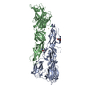
Trichoplusia ni (cabbage looper) / Strain (production host): Hi5 / References: UniProt: P17763
#2: Sugar ChemComp-NAG / 2-acetamido-2-deoxy-beta-D-glucopyranose / N-Acetylglucosamine


Type: D-saccharide, beta linking / Mass: 221.208 Da / Num. of mol.: 2
Source method: isolated from a genetically manipulated source
Formula: C8H15NO6
IdentifierTypeProgram
DGlcpNAcbCONDENSED IUPAC CARBOHYDRATE SYMBOLGMML 1.0
N-acetyl-b-D-glucopyranosamineCOMMON NAMEGMML 1.0
b-D-GlcpNAcIUPAC CARBOHYDRATE SYMBOLPDB-CARE 1.0
GlcNAcSNFG CARBOHYDRATE SYMBOLGMML 1.0
#3: Chemical
ChemComp-CD / CADMIUM ION


Mass: 112.411 Da / Num. of mol.: 4 / Source method: obtained synthetically / Formula: Cd
#4: Chemical ChemComp-CL / CHLORIDE ION / Chloride


Mass: 35.453 Da / Num. of mol.: 1 / Source method: obtained synthetically / Formula: Cl
#5: Water ChemComp-HOH / water / Water


Mass: 18.015 Da / Num. of mol.: 223 / Source method: isolated from a natural source / Formula: H2O

-
Experimental details

-
Experiment

ExperimentMethod: X-RAY DIFFRACTION / Number of used crystals: 1

-
Sample preparation

CrystalDensity Matthews: 2.67 Å3/Da / Density % sol: 53.99 %
Crystal growTemperature: 293 K / Method: vapor diffusion, hanging drop / pH: 4.5
Details: pH 4.5, VAPOR DIFFUSION, HANGING DROP, temperature 293K

-
Data collection

DiffractionMean temperature: 200 K
Diffraction sourceSource: SYNCHROTRON / Site: ALS / Beamline: 8.2.2 / Wavelength: 0.999 Å
DetectorType: ADSC QUANTUM 315 / Detector: CCD / Date: Nov 13, 2011 / Details: mirrors
RadiationMonochromator: Double crystal, Si(111) / Protocol: SINGLE WAVELENGTH / Monochromatic (M) / Laue (L): M / Scattering type: x-ray
Radiation wavelengthWavelength: 0.999 Å / Relative weight: 1
Reflection twinType: merohedral / Operator: h,-h-k,-l / Fraction: 0.5
ReflectionResolution: 2.57→50 Å / Num. all: 31819 / Num. obs: 31819 / % possible obs: 99.6 % / Observed criterion σ(F): -3 / Observed criterion σ(I): -3 / Rmerge(I) obs: 0.116
Reflection shellResolution: 2.57→2.65 Å / Redundancy: 7.3 % / Rmerge(I) obs: 0.521 / Mean I/σ(I) obs: 1.92 / Num. unique all: 3046 / % possible all: 99.1

-
Processing

Software
NameVersionClassification
HKL-2000data collection
PHENIX(phenix.refine: 1.8_1069)refinement
HKL-2000data reduction
HKL-2000data scaling
RefinementMethod to determine structure: MOLECULAR REPLACEMENT / Resolution: 2.57→28.481 Å / σ(F): 1.4 / Phase error: 20.54 / Stereochemistry target values: TWIN_LSQ_F
RfactorNum. reflection% reflectionSelection details
Rfree0.2099 1562 4.92 %RANDOM
Rwork0.1769 ---
obs0.183 31744 99.64 %-
all-31744 --
Solvent computationShrinkage radii: 0.9 Å / VDW probe radii: 1.11 Å / Solvent model: FLAT BULK SOLVENT MODEL
Refinement stepCycle: LAST / Resolution: 2.57→28.481 Å
ProteinNucleic acidLigandSolventTotal
Num. atoms5944 0 33 223 6200
Refine LS restraints
Refine-IDTypeDev idealNumber
X-RAY DIFFRACTIONf_bond_d0.0036132
X-RAY DIFFRACTIONf_angle_d0.6428299
X-RAY DIFFRACTIONf_dihedral_angle_d15.2492242
X-RAY DIFFRACTIONf_chiral_restr0.04990
X-RAY DIFFRACTIONf_plane_restr0.0021038
LS refinement shell
Resolution (Å)Rfactor RfreeNum. reflection RfreeRfactor RworkNum. reflection RworkRefine-ID% reflection obs (%)
2.5717-2.65460.34451350.25262707X-RAY DIFFRACTION94
2.6546-2.74930.24951420.21362745X-RAY DIFFRACTION95
2.7493-2.85910.23691420.20012731X-RAY DIFFRACTION95
2.8591-2.98890.23281430.18372759X-RAY DIFFRACTION95
2.9889-3.1460.21071410.17472714X-RAY DIFFRACTION95
3.146-3.34240.25221470.17462718X-RAY DIFFRACTION95
3.3424-3.59930.20171380.18142734X-RAY DIFFRACTION95
3.5993-3.95950.19051430.17282751X-RAY DIFFRACTION95
3.9595-4.52770.21481380.16062757X-RAY DIFFRACTION95
4.5277-5.68640.17951480.14832726X-RAY DIFFRACTION95
5.6864-21.3310.22171440.2092773X-RAY DIFFRACTION95

+
About Yorodumi

-
News

-
Feb 9, 2022. New format data for meta-information of EMDB entries

New format data for meta-information of EMDB entries

  • Version 3 of the EMDB header file is now the official format.
  • The previous official version 1.9 will be removed from the archive.

Related info.:EMDB header

External links:wwPDB to switch to version 3 of the EMDB data model

-
Aug 12, 2020. Covid-19 info

Covid-19 info

URL: https://pdbj.org/emnavi/covid19.php

New page: Covid-19 featured information page in EM Navigator.

Related info.:Covid-19 info / Mar 5, 2020. Novel coronavirus structure data

+
Mar 5, 2020. Novel coronavirus structure data

Novel coronavirus structure data

Related info.:Yorodumi Speices / Aug 12, 2020. Covid-19 info

External links:COVID-19 featured content - PDBj / Molecule of the Month (242):Coronavirus Proteases

+
Jan 31, 2019. EMDB accession codes are about to change! (news from PDBe EMDB page)

EMDB accession codes are about to change! (news from PDBe EMDB page)

  • The allocation of 4 digits for EMDB accession codes will soon come to an end. Whilst these codes will remain in use, new EMDB accession codes will include an additional digit and will expand incrementally as the available range of codes is exhausted. The current 4-digit format prefixed with “EMD-” (i.e. EMD-XXXX) will advance to a 5-digit format (i.e. EMD-XXXXX), and so on. It is currently estimated that the 4-digit codes will be depleted around Spring 2019, at which point the 5-digit format will come into force.
  • The EM Navigator/Yorodumi systems omit the EMD- prefix.

Related info.:Q: What is EMD? / ID/Accession-code notation in Yorodumi/EM Navigator

External links:EMDB Accession Codes are Changing Soon! / Contact to PDBj

+
Jul 12, 2017. Major update of PDB

Major update of PDB

  • wwPDB released updated PDB data conforming to the new PDBx/mmCIF dictionary.
  • This is a major update changing the version number from 4 to 5, and with Remediation, in which all the entries are updated.
  • In this update, many items about electron microscopy experimental information are reorganized (e.g. em_software).
  • Now, EM Navigator and Yorodumi are based on the updated data.

External links:wwPDB Remediation / Enriched Model Files Conforming to OneDep Data Standards Now Available in the PDB FTP Archive

-
Yorodumi

Thousand views of thousand structures

  • Yorodumi is a browser for structure data from EMDB, PDB, SASBDB, etc.
  • This page is also the successor to EM Navigator detail page, and also detail information page/front-end page for Omokage search.
  • The word "yorodu" (or yorozu) is an old Japanese word meaning "ten thousand". "mi" (miru) is to see.

Related info.:EMDB / PDB / SASBDB / Comparison of 3 databanks / Yorodumi Search / Aug 31, 2016. New EM Navigator & Yorodumi / Yorodumi Papers / Jmol/JSmol / Function and homology information / Changes in new EM Navigator and Yorodumi

Read more