[English] 日本語
Yorodumi
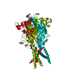
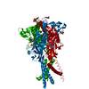
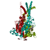
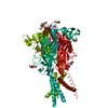
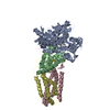
- PDB-5svj: Crystal structure of the ATP-gated human P2X3 ion channel in the ... -

+
Open data


ID or keywords:

Loading...

-
Basic information

Entry
Database: PDB / ID: 5svj
TitleCrystal structure of the ATP-gated human P2X3 ion channel in the closed, apo state
ComponentsP2X purinoceptor 3
KeywordsMEMBRANE PROTEIN / Ion Channel / Apo State
Function / homology
Function and homology information


Platelet homeostasis / purinergic nucleotide receptor activity / extracellularly ATP-gated monoatomic cation channel activity / neuromuscular synaptic transmission / Elevation of cytosolic Ca2+ levels / urinary bladder smooth muscle contraction / peristalsis / response to carbohydrate / inorganic cation transmembrane transport / cellular response to ATP ...Platelet homeostasis / purinergic nucleotide receptor activity / extracellularly ATP-gated monoatomic cation channel activity / neuromuscular synaptic transmission / Elevation of cytosolic Ca2+ levels / urinary bladder smooth muscle contraction / peristalsis / response to carbohydrate / inorganic cation transmembrane transport / cellular response to ATP / positive regulation of calcium ion transport into cytosol / behavioral response to pain / protein homotrimerization / positive regulation of calcium-mediated signaling / response to mechanical stimulus / hippocampal mossy fiber to CA3 synapse / response to cold / establishment of localization in cell / calcium ion transmembrane transport / regulation of synaptic plasticity / Schaffer collateral - CA1 synapse / sensory perception of taste / response to heat / postsynapse / response to hypoxia / receptor complex / axon / signal transduction / ATP binding / plasma membrane
Similarity search - Function
P2X3 purinoceptor / atp-gated p2x4 ion channel / atp-gated p2x4 ion channel fold / atp-gated p2x4 ion channel domain / ATP P2X receptor / ATP P2X receptors signature. / P2X purinoreceptor / P2X purinoreceptor extracellular domain superfamily / Helix Hairpins / Sandwich ...P2X3 purinoceptor / atp-gated p2x4 ion channel / atp-gated p2x4 ion channel fold / atp-gated p2x4 ion channel domain / ATP P2X receptor / ATP P2X receptors signature. / P2X purinoreceptor / P2X purinoreceptor extracellular domain superfamily / Helix Hairpins / Sandwich / Orthogonal Bundle / Mainly Beta / Mainly Alpha
Similarity search - Domain/homology
Biological speciesHomo sapiens (human)
MethodX-RAY DIFFRACTION / SYNCHROTRON / MOLECULAR REPLACEMENT / Resolution: 2.984 Å
AuthorsMansoor, S.E. / Lu, W. / Oosterheert, W. / Shekhar, M. / Tajkhorshid, E. / Gouaux, E.
Funding support United States, 2items
OrganizationGrant numberCountry
National Institutes of Health/National Institute of General Medical Sciences (NIH/NIGMS)R01GM100400 United States
National Institutes of Health/National Institute of General Medical Sciences (NIH/NIGMS)5F32GM108391 United States
CitationJournal: Nature / Year: 2016
Title: X-ray structures define human P2X3 receptor gating cycle and antagonist action.
Authors: Mansoor, S.E. / Lu, W. / Oosterheert, W. / Shekhar, M. / Tajkhorshid, E. / Gouaux, E.
History
DepositionAug 6, 2016Deposition site: RCSB / Processing site: RCSB
Revision 1.0Sep 28, 2016Provider: repository / Type: Initial release
Revision 1.1Oct 19, 2016Group: Database references
Revision 1.2Sep 20, 2017Group: Author supporting evidence / Database references / Derived calculations
Category: citation / pdbx_audit_support / pdbx_struct_oper_list
Item: _citation.journal_id_CSD / _pdbx_audit_support.funding_organization / _pdbx_struct_oper_list.symmetry_operation
Revision 1.3Dec 25, 2019Group: Author supporting evidence / Category: pdbx_audit_support / Item: _pdbx_audit_support.funding_organization
Revision 1.4Jul 29, 2020Group: Data collection / Derived calculations / Structure summary
Category: chem_comp / entity ...chem_comp / entity / pdbx_chem_comp_identifier / pdbx_entity_nonpoly / struct_conn / struct_site / struct_site_gen
Item: _chem_comp.name / _chem_comp.type ..._chem_comp.name / _chem_comp.type / _entity.pdbx_description / _pdbx_entity_nonpoly.name / _struct_conn.pdbx_role
Description: Carbohydrate remediation / Provider: repository / Type: Remediation

-
Structure visualization

Structure viewerMolecule:
MolmilJmol/JSmol

Downloads & links

-
Assembly

Deposited unit
A: P2X purinoceptor 3
hetero molecules


Theoretical massNumber of molelcules
Total (without water)41,7138
Polymers40,7461
Non-polymers9677
Water46826
1
A: P2X purinoceptor 3
hetero molecules

A: P2X purinoceptor 3
hetero molecules

A: P2X purinoceptor 3
hetero molecules


Theoretical massNumber of molelcules
Total (without water)125,13924
Polymers122,2383
Non-polymers2,90221
Water543
TypeNameSymmetry operationNumber
identity operation1_555x,y,z1
crystal symmetry operation2_555-y,x-y,z1
crystal symmetry operation3_555-x+y,-x,z1
Buried area16090 Å2
ΔGint-13 kcal/mol
Surface area44340 Å2
MethodPISA
Unit cell
Length a, b, c (Å)120.170, 120.170, 236.580
Angle α, β, γ (deg.)90.00, 90.00, 120.00
Int Tables number155
Space group name H-MH32
Components on special symmetry positions
IDModelComponents
11A-405-

NA

-
Components

-
Protein / Sugars , 2 types, 4 molecules A

#1: Protein P2X purinoceptor 3 / P2X3 / ATP receptor / Purinergic receptor


Mass: 40745.918 Da / Num. of mol.: 1
Source method: isolated from a genetically manipulated source
Source: (gene. exp.) Homo sapiens (human) / Gene: P2RX3 / Cell line (production host): HEK293 / Production host: Homo sapiens (human) / References: UniProt: P56373
#2: Sugar ChemComp-NAG / 2-acetamido-2-deoxy-beta-D-glucopyranose / N-Acetylglucosamine


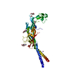
Type: D-saccharide, beta linking / Mass: 221.208 Da / Num. of mol.: 3
Source method: isolated from a genetically manipulated source
Formula: C8H15NO6
IdentifierTypeProgram
DGlcpNAcbCONDENSED IUPAC CARBOHYDRATE SYMBOLGMML 1.0
N-acetyl-b-D-glucopyranosamineCOMMON NAMEGMML 1.0
b-D-GlcpNAcIUPAC CARBOHYDRATE SYMBOLPDB-CARE 1.0
GlcNAcSNFG CARBOHYDRATE SYMBOLGMML 1.0

-
Non-polymers , 5 types, 30 molecules

#3: Chemical ChemComp-MG / MAGNESIUM ION


Mass: 24.305 Da / Num. of mol.: 1 / Source method: obtained synthetically / Formula: Mg
#4: Chemical ChemComp-NA / SODIUM ION


Mass: 22.990 Da / Num. of mol.: 1 / Source method: obtained synthetically / Formula: Na
#5: Chemical ChemComp-PG4 / TETRAETHYLENE GLYCOL / Polyethylene glycol


Mass: 194.226 Da / Num. of mol.: 1 / Source method: obtained synthetically / Formula: C8H18O5 / Comment: precipitant*YM
#6: Chemical ChemComp-EDO / 1,2-ETHANEDIOL / ETHYLENE GLYCOL / Ethylene glycol


Mass: 62.068 Da / Num. of mol.: 1 / Source method: obtained synthetically / Formula: C2H6O2
#7: Water ChemComp-HOH / water / Water


Mass: 18.015 Da / Num. of mol.: 26 / Source method: isolated from a natural source / Formula: H2O

-
Experimental details

-
Experiment

ExperimentMethod: X-RAY DIFFRACTION / Number of used crystals: 1

-
Sample preparation

CrystalDensity Matthews: 4.01 Å3/Da / Density % sol: 69.32 %
Crystal growTemperature: 277.15 K / Method: vapor diffusion, hanging drop / Details: 25% PEG 400, 100 mM MES, pH 6.85, 50 mM MgCl2

-
Data collection

DiffractionMean temperature: 100 K
Diffraction sourceSource: SYNCHROTRON / Site: ALS / Beamline: 5.0.2 / Wavelength: 1 Å
DetectorType: ADSC QUANTUM 315 / Detector: CCD / Date: Sep 20, 2015
RadiationProtocol: SINGLE WAVELENGTH / Monochromatic (M) / Laue (L): M / Scattering type: x-ray
Radiation wavelengthWavelength: 1 Å / Relative weight: 1
ReflectionResolution: 2.98→50 Å / Num. obs: 13067 / % possible obs: 95.4 % / Redundancy: 4.12 % / Net I/σ(I): 18.34

-
Processing

Software
NameVersionClassification
PHENIX(1.10_2142: ???)refinement
XDSdata reduction
XDSdata scaling
PHENIXphasing
RefinementMethod to determine structure: MOLECULAR REPLACEMENT / Resolution: 2.984→39.068 Å / SU ML: 0.41 / Cross valid method: FREE R-VALUE / σ(F): 1.34 / Phase error: 29.18 / Stereochemistry target values: ML
RfactorNum. reflection% reflection
Rfree0.2591 638 4.88 %
Rwork0.2125 --
obs0.2147 13062 95.43 %
Solvent computationShrinkage radii: 0.9 Å / VDW probe radii: 1.11 Å / Solvent model: FLAT BULK SOLVENT MODEL
Refinement stepCycle: LAST / Resolution: 2.984→39.068 Å
ProteinNucleic acidLigandSolventTotal
Num. atoms2447 0 61 26 2534
Refine LS restraints
Refine-IDTypeDev idealNumber
X-RAY DIFFRACTIONf_bond_d0.0032567
X-RAY DIFFRACTIONf_angle_d0.7723493
X-RAY DIFFRACTIONf_dihedral_angle_d16.5511511
X-RAY DIFFRACTIONf_chiral_restr0.05407
X-RAY DIFFRACTIONf_plane_restr0.004441
LS refinement shell
Resolution (Å)Rfactor RfreeNum. reflection RfreeRfactor RworkNum. reflection RworkRefine-ID% reflection obs (%)
2.9841-3.21440.36611170.31612142X-RAY DIFFRACTION84
3.2144-3.53760.28651400.24772549X-RAY DIFFRACTION99
3.5376-4.04910.23661470.21092539X-RAY DIFFRACTION99
4.0491-5.09960.23861220.17962573X-RAY DIFFRACTION99
5.0996-39.07090.25751120.21112621X-RAY DIFFRACTION96

+
About Yorodumi

-
News

-
Feb 9, 2022. New format data for meta-information of EMDB entries

New format data for meta-information of EMDB entries

  • Version 3 of the EMDB header file is now the official format.
  • The previous official version 1.9 will be removed from the archive.

Related info.:EMDB header

External links:wwPDB to switch to version 3 of the EMDB data model

-
Aug 12, 2020. Covid-19 info

Covid-19 info

URL: https://pdbj.org/emnavi/covid19.php

New page: Covid-19 featured information page in EM Navigator.

Related info.:Covid-19 info / Mar 5, 2020. Novel coronavirus structure data

+
Mar 5, 2020. Novel coronavirus structure data

Novel coronavirus structure data

Related info.:Yorodumi Speices / Aug 12, 2020. Covid-19 info

External links:COVID-19 featured content - PDBj / Molecule of the Month (242):Coronavirus Proteases

+
Jan 31, 2019. EMDB accession codes are about to change! (news from PDBe EMDB page)

EMDB accession codes are about to change! (news from PDBe EMDB page)

  • The allocation of 4 digits for EMDB accession codes will soon come to an end. Whilst these codes will remain in use, new EMDB accession codes will include an additional digit and will expand incrementally as the available range of codes is exhausted. The current 4-digit format prefixed with “EMD-” (i.e. EMD-XXXX) will advance to a 5-digit format (i.e. EMD-XXXXX), and so on. It is currently estimated that the 4-digit codes will be depleted around Spring 2019, at which point the 5-digit format will come into force.
  • The EM Navigator/Yorodumi systems omit the EMD- prefix.

Related info.:Q: What is EMD? / ID/Accession-code notation in Yorodumi/EM Navigator

External links:EMDB Accession Codes are Changing Soon! / Contact to PDBj

+
Jul 12, 2017. Major update of PDB

Major update of PDB

  • wwPDB released updated PDB data conforming to the new PDBx/mmCIF dictionary.
  • This is a major update changing the version number from 4 to 5, and with Remediation, in which all the entries are updated.
  • In this update, many items about electron microscopy experimental information are reorganized (e.g. em_software).
  • Now, EM Navigator and Yorodumi are based on the updated data.

External links:wwPDB Remediation / Enriched Model Files Conforming to OneDep Data Standards Now Available in the PDB FTP Archive

-
Yorodumi

Thousand views of thousand structures

  • Yorodumi is a browser for structure data from EMDB, PDB, SASBDB, etc.
  • This page is also the successor to EM Navigator detail page, and also detail information page/front-end page for Omokage search.
  • The word "yorodu" (or yorozu) is an old Japanese word meaning "ten thousand". "mi" (miru) is to see.

Related info.:EMDB / PDB / SASBDB / Comparison of 3 databanks / Yorodumi Search / Aug 31, 2016. New EM Navigator & Yorodumi / Yorodumi Papers / Jmol/JSmol / Function and homology information / Changes in new EM Navigator and Yorodumi

Read more